Monday, November 14, 2016

Deep Sulcus Sign - Pneumothorax

Pneumothorax simply refers to the presence of air in the pleural space. When this collection of air enlarges, it leads to the compression of mediastinal structures causing life-threatening tension pneumothorax. Pneumothoraces can be divided into three categories:
  1. Primary spontaneous: no underlying lung disease (Connective tissue disorders)
  2. Secondary spontaneous: underlying lung disease is present (COPD)
  3. Iatrogenic/traumatic (trauma, biopsy, barotrauma)

Diagnosing Pneumothorax: 
  • History and Physical Examination
  • Bedside Lung USG
  • CXR
  • CT scan

USG - Check out this link to learn more about USG
M-mode can be used to determine movement of lung within the rib-interspace. Small pneumothoraces are best appreciated anteriorly in the supine position (gas rises) whereas large pneumothoraces are appreciated laterally in the mid-axillary line.


CXR in Pneumothorax 

A classical CXR shows: 
  • Visible visceral pleural edge seen as a very thin, sharp white line
  • No lung markings are seen peripheral to this line i.e. high Radiolucency 
  • Peripheral space is radiolucent compared to adjacent lung 
  • Completely collapse of lung
  • Mediastinum shift is seen only in tension pneumothorax
  • Subcutaneous Emphysema
On an erect chest radiograph, a pneumothorax will usually be apical. It can be challenging to visualise if it is small. Look carefully in the region above the line of the clavicle.


See pneumothorax demarcated on the second Chest X

 On your left, see the inspiratory film with PTx marked on the Right lung with yellow arrows. On your right, see the Expiratory film which shrinks the lungs and makes the Pneumothorax more obvious. Note the collapsed Right lung in this image. 


The Deep Sulcus Sign (CXR)
Pneumothorax can sometimes become a difficult diagnosis. Patients don't read textbooks and may not give you a classic presentation such as obvious hypoxia and absent breath sounds. 
When USG is available, start with a quick lung USG but it can be difficult to comment on breath sounds in a patient who is bleeding from nostrils and breathing at 40/min with broken ribs. USG with M-Mode still is your best bet here. CXR cannot be used to rule out a pneumothorax. Another issue is that ED patients are usually supine and a small pneumothorax float anteriorly in the supine position, and if it is not big enough to wrap around the lateral edge of the lung, it may be missed. So we need to be aware of subtle signs on a CXR. The deep sulcus sign is one of the more subtle signs. 
The deep sulcus sign is a dark lateral sulcus where the chest wall meets the diaphragm. The amount of lung in this area is less, so a small amount of air will tend to darken the area making it more prominent. 


If you see a deep sulcus sign on the CXR, strongly consider pneumothorax. If the patient is unstable, do needle decompression and insert with a chest tube. Aldo remember that a CXR always underestimates the true size of the pneumothorax. 
Patients with COPD may exhibit deepened lateral costophrenic angles due to hyperaeration of the lungs and cause a false deep sulcus sign.

CT scan - CT shows even the smallest of Pnemothoraces. 


I age from trauma.oh
Right tension pneumothorax - CT scan
The mediastinum is shifted to the left and the right lung compressed posteriorly

Take Home:
  • PTx Diagnosis - Begin with History and Clinical exam followed by bedside USG - look for lung sliding (Lung sliding is Normal)
  • Scrutinise the CXR and look for deep sulcus sign but beware of false deep sulcus in COPDs.
  • CT only if high suspicion of Pneumothorax with equivocal USG and CXR 


Author:

              
     Lakshay Chanana
     
     Speciality Doctor
     Northwick Park Hospital
     Department of Emergency Medicine
     England

     @EMDidactic


Monday, November 7, 2016

Chasing Chest Pains - Risk Stratification and Non Specific Troponinemia

Chest pain is caused by a myriad of causes ranging from benign to life threatening, some of which can cause death within minutes or hours. While evaluating chest pain, ACS is always high up on our list of differential diagnosis and as Emergency Physicians, it is our responsibility to not only robustly identify ACS and but also avoid needless investigations and unnecessary admissions for those who can be safely discharged from the ED after risk stratification.


Here is an overview of ACS with breakdown of terminologies, key points about history/physical exam and biomarkers and specifics about what to do with a low risk ACS patient:

Classical ACS presents with:
  •             Heavy, aching or tight
  •             Central chest or left sided
  •             Not related to respiration or movement
  •             May radiate to arms, neck, or jaw


You will often see patients who have one or a few of these features but end up having a completely negative work up for ACS. Remember, the history is helpful only to risk stratify – not to confirm your diagnosis. Everyone perceives pain in a different way but history is the first step during evaluation and risk stratification. 

Atypical ACS
Atypical presentations of ACS are common, occurring in up to 1/3rd of patients, mostly in the elderly, diabetics and women. Advanced age, co-morbid factors, delay in diagnosis lead to the increased mortality in these populations.


Things you must ask/look for:

  • Radiation to both arms – Likely  ACS
  • Radiation to left arm – Likely ACS
  • Nausea / Vomiting – Likely ACS
  • SOB on exertion – Likely ACS
  • Associated with Sweating – Likely ACS
  • Hypotension – Likely ACS
  • S3 – Likely ACS
  • Describes as previous angina - Likely ACS


  • Pleuritic/ Positional/ Sharp Pain – Unlikely ACS (not impossible)
  • Tender on Palpation – Unlikely ACS (not impossible)


A good history helps in risk stratification. Don’t rule out ACS just based on the history alone. With the slightest of concern, get an ECG.


Risk Factors
Risk factors once again help us to risk stratify but just based on the absence of risk factors you cannot rule out ACS. Get worried if the history is concerning even if there are no risk factors at all. The next step is ECG.


And what if the history is concerning but ECG is normal?
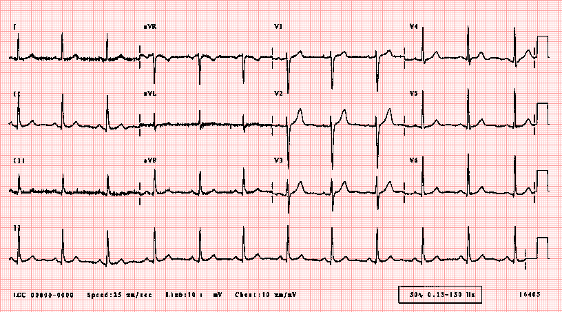
An ECG showing ST depressions, TWI or STE is obviously concerning. Patients who present with chest pain with suspected cardiac ischemia based on the history but normal ECG should still undergo further diagnostic testing with cardiac markers before they can be confidently assigned to a low risk group.


What if the history is concerning with ongoing ischemic symptoms, but ECG is normal and troponin is not elevated?
This sounds very much like Unstable Angina. DO NOT SEND ANYONE HOME WITH CONCERNING AND PERSISTING SYMPTOMS DESPITE NEGATIVE TROPONINS.

Unstable Angina can occur when you are resting, sleeping, or during  little physical exertion. The pain may last longer than stable angina and rest or anti-ischemic medications usually do not help relieve it. USA can have an ischemic or normal ECG but should always have negative troponins by definition.

In contrast, Stable Angina is very predictable with Chest Pain on exertion that gets better on resting. Stable Angina us also relieved with anti-ischemic medications.


Beware of the Non-Specific Troponinemia AKA Troponinitis!

Troponins are the preferred and recommended markers of myocardial necrosis. Read more about troponins here. But the new generation hs troponins are extremely sensitive and thus less specific i.e you might end up getting a false positive elevated troponin leading to unnecessary admissions and work ups.  So if history is not suggestive of ACS but troponin is elevated – get a few more ECGs but do think of other possible causes of an elevated troponin such as:


With hs Troponins, we are now able to pick up even the minimal level of troponins in a patient’s system and thus those who were diagnosed as Unstable Anginas (Ischemic Chest Pain/ECG but normal troponin levels) a decade back are now labeled as NSTEMIs (Ischemic Chest Pain/ECG but a raised troponin). At this moment, although it is controversial but some experts do believe that Unstable Angina does not even exist anymore in this era of highly sensitive troponins.


ACS includes STEMI, NSTEMI, USA (not stable angina)
Patients with STEMI do not require troponin since their initial treatment is determined by their clinical presentation and ECG findings. Patients with STEMI are identified quickly, assigned a high risk category and have a well-defined treatment strategy (ie. urgent reperfusion with PCI or thrombolytics).


When does NSTEMI need immediate cathlab:
The ACC/AHA guidelines for NSTEMI recommend < 2 hour cath for:
  1. Refractory ischemia
  2. Ischemia with hemodynamic or electrical instability


If you are worried about a patient, get serial ECGs, send troponins and involve cardiology at the earliest.


Disposition of a Low Risk Patient – Slightly concerning history but non-ischemic ECG and negative enzymes. 

Here we are specifically talking about Unstable Anginas which can be further divided into two groups i.e negative troponin with ischemic ECG and negative troponin with a non-ischemic ECG. 

Current data shows that if patients have negative troponins with non-ischemic ECG, then prognosis is not bad even if they are diagnosed with unstable angina.  If they have unstable angina with an ischemic EKG, then they have a worse prognosis.

Note - if you see an Ischemic ECG – Get worried even when if the enzymes are normal

Low risk unstable angina with negative troponins can have:
Shared Medical Decision Making -  Do serial troponins and serial ECGs. Current evidence suggests a repeat troponin at hour 3 from initial EKG reduces potential miss rate from 1.7% to <1% at 30 days. Let them make this decision - ask them if they would want to get admitted or if they are happy to follow up as an out-patient with a week. 


Use Clinical Decision Making rules such as HEART/GRACE score to further risk stratify them and most important - Document medical decision-making and Clinical Decision rules  in the patient's record.


Read more on HEART SCORE on REBELEM.


Take Home:
  • A good history helps in risk stratification. Don’t rule out ACS just based on the history alone. With the slightest of concern, get an ECG.
  • It is okay to send troponins on your patients if you have some degree of concern but If there are no concerns eat all, then do not send troponins.
  • Patients who present with chest pain with suspected cardiac ischaemia based on the history but normal ECG should still undergo further diagnostic testing.
  • USA can have an ischemic or normal ECG but should always have negative troponins by definition.
  • Low Risk - Do Serial ECGs, Shared Decision Making, Clinical Decision Making Rules to further risk stratify and DOCUMENT the decision making in the medical record. 


Further Reading:
  • http://hqmeded-ecg.blogspot.co.uk/2014/04/unstable-angina-dr-braunwald-asks-if-it.html
  • http://hqmeded-ecg.blogspot.co.uk/2015/06/unstable-angina-still-exists-beware.html
  • https://blog.essentialsofem.com/2016/02/25/low-risk-chest-pain-adp-showdown-using-timi-vs-heart-pt-1-of-3-timi/


Author:

              
     Lakshay Chanana
     
     Speciality Doctor
     Northwick Park Hospital
     Department of Emergency Medicine
     England

     @EMDidactic


Monday, October 31, 2016

Is troponin really becoming a lousy test?

Just a quick reminder this week regarding the use of troponin. This is one of the most abused tests in Emergency Departments these days.  The hs Troponins seems to be overtly sensitive that might lead to unnecessary admissions. Here are a few caveats with troponin:

1. Do a history first, followed by a physical exam and then ECG. Only after that, send a troponin if you are concerned about the possibility of ACS. 

DO NOT ORDER TROPONIN FOR EVERYONE WALKING IN WITH CHEST PAIN

2. Remember that there a  plenty of other reasons to have a positive troponin besides ACS. To name a few - PE, Heart Failure, Sepsis, Renal Failure, Stroke, Tachycardia, Infiltrative Heart Disease, Myopericarditis.

A RAISED TROPONIN DOES NOT ALWAYS EQUAL ACS. TROPONIN IS A MARKER OF ANY MYOCARDIAL INJURY, NOT JUST ISCHEMIA. 

3. STEMI does not need a troponin. Focus on rapid Reperfusion (Thrombolytics or PCI). Troponin is done to differentiate between NSTEMI and USA (Unstable Angina).


4. Discharging someone with a single negative troponin is high risk business (unless it was drawn 6 hours after the onset of symptoms). Whenever possible, get two serial troponins to see if there is any significant rise form the baseline. However, serial sampling of hs troponin at 0 and 2 hours can safely rule-out of STEMI and NSTEMI. 

In case, you happen to discharge home a patient with two negative troponins, document why are you doing that because it can still be Unstable Angina. 

5. Troponin might take 6-12 hours to rise from the onset of symptoms and stays elevated upto 2 weeks. Document your concerns while sending these cardiac biomarkers.


Attorneys focus on the documentation of HPI, documentation of ECG findings and Medical Decision Making with appropriate reasoning before settling the case. We are going to cover these bits next week. 


BOTTOMLINE: I think troponin is still a great test, if done with caveats. However, over the last two decades, it has become extremely sensitive and thus non-specific (like d-dimer for PE). 


Further Reading: 


Author:

              
     Lakshay Chanana
     
     Speciality Doctor
     Northwick Park Hospital
     Department of Emergency Medicine
     England
     @EMDidactic


Monday, October 24, 2016

Hundred thousand views

A big thanks to each and everyone of you out there who supported the existence of this blog. To be honest, being the first one from India was quite intimidating but I am pleased to know that the blog has recently crossed the milestone of 100,000 hits in less than 2 years. Things were pretty rough when I began this endeavour in January 2015. There were critics and skeptics who questioned every bit of it. Fortunately, we have come a long way since then and now more and more people are open to Free Open Access Medical Education.


A special mention about all the contributors for taking out time from their busy schedules. It was not possible to reach this far without your support. Thanks a ton guys!!

  • Akshay Kumar
  • Apoorva Chandra
  • Azharullah Khan
  • Kritika Atrey 
  • Liaquat Roopesh
  • Nikhil Tambe
  • Sagar Galwankar

I usually post a topic on every Monday and it would not have been possible without the support of Karishma, my wife. She has sacrificed numerous weekends and whenever it was about the blog - there were no questions asked! She has been a huge help and also one of the major critics of this blog. 


Over the course of last couple of years, many folks asked me about the blog logo. Here is what it signifies..


The logo took its inspiration from Emergency Medical Services. It is comprised of the star of life - a symbol most commonly associated with EMS. The six branches of the star symbolise 
  1. Early Detection
  2. Early Reporting
  3. Early Response
  4. On Scene Care
  5. Care in Transit
  6. Transfer to Definitive care
The snake emblem is the Rod of Asclepius is the symbol of medical care, named for the Greek mythological figure Asclepius, who was said to have possessed healing power.

The numerous yellow colored light bulbs within and around the star represent dandelions of ideas. The ideas do have their origin and base within Emergency Medicine but they travel out to other zones to disperse and germinate as novel thoughts and innovations. 

Credits to Kritika Atrey for crafting this logo for the blog. 


And for those of you who read and COMMENT on the posts, just to let you know that you  people make my day. A word of appreciation means a lot and keeps me going. Your intrigue questions, thoughts and comments always open an opportunity for discussions.


Lastly, this note is incomplete without acknowledging the contribution of "The Teaching Course" which played an indispensable role behind the inception of this blog. This course was phenomenal and it turned out to be a life-changing experience for me. It was absolutely worth travelling over 8000miles to be there. I owe it to these people.


Click here to know more about the teaching course. 


Author:


     Lakshay Chanana
     
     Speciality Doctor
     Northwick Park Hospital
     Department of Emergency Medicine
     England

     @EMDidactic