Monday, October 30, 2017

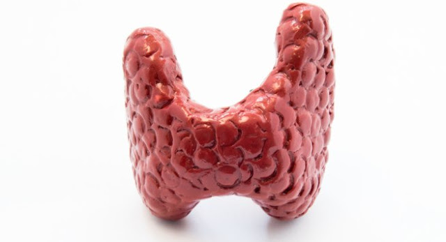
Thyroid Storm

Thyroid hormone exists in two forms: thyroxine and triiodothyronine. The ratio of thyroxine to triiodothyronine released in the blood is about 20:1 and peripherally, thyroxine is converted to the active triiodothyronine (T3), which is three to four times more potent than thyroxine.

Basic Terminologies
  • Primary hyperthyroidism is caused by the excess production of thyroid hormones from the thyroid glands. 
  • Secondary hyperthyroidism is caused by the excess production of thyroid-releasing hormones (Hypothalamus) or thyroid-stimulating hormones (Pituitary).

  • Hyperthyroidism refers to excess circulating hormone resulting only from thyroid gland hyper function. Most commonly cause by Graves' Disease. 
  • Thyrotoxicosis refers to excess circulating thyroid hormone originating from any cause. 
  • Thyroid storm is an extreme manifestation of thyrotoxicosis. It presents as an acute life-threatening hypermetabolic state caused either by excessive release of thyroid hormones or due to altered peripheral response to thyroid hormone following a precipitating event. 

Causes of Hyperthyroidism
  • Graves' Disease
  • Toxic Goitre
  • Thyroiditis (Viral, Radiation)
  • Hashimoto's
  • Secondary (Pituitary or Hypothalamus related)
  • Thyrotoxicosis Factitia
  • Drug Induced (Amiodarone, Interleukin 2)
  • Metastatic (Struma Ovarii)
  • Hydatidiform Mole

Precipitants of Thyroid Storm
  • Trauma
  • Heat related Illness
  • Recreational Drug Use
  • Psychosis
  • Stress
  • Infection
  • Iodinated Contrasts
  • ACS
  • DKA/HHS
  • Thyroxine Overdose
Consider thyroid Strom in any patient presenting with fear, tachycardia and altered mental status. Use Burch-Wartofsky scale to gauge your suspicion and diagnosis. 


Management
Following the order of treatment is of utmost importance here. Inhibition of thyroid gland synthesis of new thyroid hormone with a thionamide (Methimazole or PTU) must be initiated before iodine therapy.


1. Supportive care 
  • ABC
  • Cardiac Monitor and IV accsess.
  • Fluids, Maintenance of Electrolytes and Glucose. Cooling for hyperthermia. 
  • Add antibiotics as infection is a known precipitant and hard to distinguish in ED
  • Consider Cholestyramine to decrease the reabsorption of thyroid hormone from the enterohepatic circulation. In thyrotoxicosis, there is increased enterohepatic circulation of thyroid hormone. 

2. Inhibition of new hormone synthesis
Thionamides: Methimazole or PropylThioUracil. Thionamides decrease the synthesis of new hormone production. 

Methimazole: 40 to 100 milligrams PO as loading dose then 20 milligrams q4h


Propylthiouracil: Load with 600 to 1000 mg PO followed by 200 to 250 milligrams every 4 hours. PTU is hepatotoxic but in addition to decreasing the synthesis of new hormone production, it also blocks the peripheral conversion of thyroxine to triiodothyronine. 


Use PTU in first trimester and Methimazole in second and third trimester. 

 3. Inhibition of thyroid hormone release
Potassium iodide can be given to stop thyroid hormone release. PTU or Methimazole must be started first and Iodine therapy should be given at least 1 hour later. Iodine therapy blocks the release of pressured hormone. Start with 8 to 10 drops initially. 


Iodine-containing solutions should not be given to patients with iodine overload or iodine-induced hyperthyroidism or amiodarone-induced thyrotoxicosis. Lithium or potassium perchlorate should be used instead. 

 4. Peripheral β-adrenergic receptor blockade

Propranolol can be given IV in slow 1- to 2-milligram boluses q5-10min. Orally, propranolol therapy usually begins at 20 to 120 milligrams per dose.

 5. Preventing peripheral conversion of thyroxine to triiodothyronine 
The peripheral conversion of thyroxine to triiodothyronine is blocked by propylthiouracil, propranolol, and glucocorticoid. Glucocorticoids are essential in treatment since blockade produced by propylthiouracil and propranolol is not significant. . Glucocorticoid also treat underlying relative adrenal insufficiency. 

6. Find and treat the precipitation event (Sepsis, DKA, ACS)
7. Definitive Treatment - Radioactive Iodine or Surgery 


Other potential considerations:
  • Direct removal of thyroid hormone with plasma exchange
  • Use of Potassium Perchlorate in Amiodarone induced thyrotoxicosis: Potassium perchlorate  interferes with the production of new hormones 
  • Lithium: Used in cases of hypersensitivity to iodine. Lithium inhibits thyroid hormone release from thyroid gland. Typical dosing in thyroid storm is 300 milligrams every 8 hours. Monitor levels to avoid toxicity. 
  • Peripheral beta blockade: Reserpine or Guanethidine can be used, if there is a contraindication for BB use. These agents do not block beta receptors and interfere with catecholamine function (by depleting stores and blocking release)

Take Home:
  • Consider Thyroid Strom in any patient presenting with fever, tachycardia and Altered Mental Status.
  • PTU or Methimazole must be started first and Iodine therapy should be given at least 1 hour later.
  • Manage with beta blockers, PTU/Methimazole, Steroids and Potassium iodide
  • Identify and treat the precipitation cause 

Further Reading
Posted by:

              
     Lakshay Chanana
     
     Speciality Doctor
     Northwick Park Hospital
     Department of Emergency Medicine
     England
     @EMDidactic


Sunday, October 22, 2017

The patient with Long lie - Rhabdomyolysis

Skeletal muscle injury caused by any mechanism (direct, genetic, biochemical) is termed as rhabdomyolysis. Injured muscle fibers release cellular contents into the circulation leading to myoglobinuria. Besides myoglobin, cellular contents also include enzymes suck as creatine kinase (CK), aldolase, LDH, AST, and potassium. Eventually, the Na+K+ATPase pump and Ca transport takes the hit resulting in increased intracellular calcium and muscle injury. Calcium also activates intracellular enzymes including proteases inducing production of free oxygen radicals. 


Pathophysiology of Rhabdomyolysis

Potential causes of rhabdomyolysis include:


  • Alcohol (Prolonged immobilisation and direct alcohol related toxicity, Low K/Mg/PO4 also predispose)
  • Recreational Drug use (Cocaine, Amphetamines, Opoids)
  • Medications (Statins, TCAs, SSRIs, Lithium, Steroids, MAOi )
  • Myopathies(Dermatomyositis, Polymyositis)
  • Trauma (Electricity, Crush Injury)
  • Seizures
  • Prolonged Immobility (Post Falls in Elderly)
  • Infection (Bacterial and Viral)
  • Strenuous physical activity
  • Heat-related illness. 


Inherited metabolic disorders should be suspected with recurrent episodes of rhabdomyolysis


Clinical Presentation
  • Acute onset muscle aches
  • Muscle stiffness and weakness
  • Fever
  • Dark coloured urine. 
  • Nausea, vomiting and tachycardia 
  • Tenderness Muscles 

Examination may be completely normal. Therefore, look for cues in the history and work up based on that.


Diagnosis

Raised CK is a marker of muscle injury. The degree of elevation correlates with the amount of damage but it is unrelated with the morbidity. A rise in CK >5 times than normal is considered as significant. CK levels begin to rise 2 to 12 hours after the onset of injury, and peak within 24 to 72 hours.


Pattern of Myoglobin and CK rise during Rhabdomyolysis


Myoglobin elevation actually occurs before CK elevation but it rapidly cleared from the plasma. It enters urine only when the plasma concentration is significantly elevated. Because myoglobin contains heme, dipstick tests may turn positive as then test cannot differentiate among hemoglobin, myoglobin, and red blood cells. Therefore, suspect myoglobinuria when the urine dipstick test is positive for blood but no red blood cells are present on microscopic examination. 


Myoglobin levels may return to normal within 1 to 6 hours after the onset of muscle necrosis, therefore absence of an elevated serum myoglobin level or of myoglobinuria does not exclude the diagnosis. 

Due to cellular injury, serum electrolyte disturbances are seen commonly (Low Ca, High K/Uric Acid/Phosphorus). Renal Function tests reflect the extent of kidney injury. 


Management

  • Treat the underlying cause
  • Aggressive Fluid Resuscitation (Normal Saline)
  • Target Urine Output is 200-300ml/hr (catheterise to check output in critically ill)
  • Treat Hyperkalemia with usual measures
  • Avoid Nephrotoxic Medications 
  • Renal Replacement therapy
  • Consider mannitol, especially if compartment syndrome - Low grade evidence 

As usual - Bicarbonate is recommended by some but without any definite evidence base. 


The idea of urine arlkalinization stems from the fact that myoglobin precipitation is increased in acidic urine. However, in clinical studies - urine alkalinization has not been shown to impact outcomes. 


Compartment syndrome and peripheral nerve injury are potential mechanical complications of Rhabdomyolysis. Rarely, thromboplastin release from damaged muscle leads to consumptive coagulopathy and DIC. 


Take Home

  • Early recognition and prompt initiation of treatment is the key
  • Pick cues from the history as examinations findings can be subtle or absent
  • Watch for the complications (DIC, Compartment Syndrome)


References:
  1. Parekh R, Care DA, Tainter CR. Rhabdomyolysis: advances in diagnosis and treatment. Emergency medicine practice. 2012 Mar;14(3):1-5.
  2. Malinoski DJ, Slater MS, Mullins RJ. Crush injury and rhabdomyolysis. Critical care clinics. 2004 Jan 1;20(1):171-92.
  3. Giannoglou GD, Chatzizisis YS, Misirli G. The syndrome of rhabdomyolysis: pathophysiology and diagnosis. European journal of internal medicine. 2007 Mar 31;18(2):90-100.
  4. ThomasR:Towardsevidence-basedemergencymedicine: bestBETS from the Manchester Royal Infirmary. Emerg Med J 27: 305, 2010. 
  5. Dalakas MC: Toxic and drug-induced myopathies. J Neurol Neurosurg Psychiatry 80: 832, 2009.
  6. Clarkson PM, Eichner ER: Exertional rhabdomyolysis: does elevated blood creatine kinase foretell renal failure? Curr Sports Med Rep 5: 57, 2006. [PMID: 16529674]
  7. Vanholder R, Sever MS, Ekrem E, et al: Rhabdomyolysis. J Am Soc Nephrol 11: 1553, 2000. 

Posted by:

              
     Lakshay Chanana
     
     Speciality Doctor
     Northwick Park Hospital
     Department of Emergency Medicine
     England

     @EMDidactic


Monday, October 16, 2017

Don't be critical about "FRCEM Critical Appraisal"

The September 2017 diet FRCEM critical appraisal results were released last week and passing rates remained close to 60%. I took this exam in September diet along with some of my other colleagues. 


While preparing for this exam, we often used to ponder - Why are we doing this? But as our preparation went on, we realized that “Critical Appraisal” is one of the most essential aspects of modern day medicine. As exam-going candidates, like everyone else we had a lot of doubts and concerns about the pattern, how to start preparation and we found that there is a ton of stuff on MRCEM Part A, B and C prep but there was not much about FRCEM Final set of exams. 


I am going to address few BIG questions regarding FRCEM Critical Appraisal here:


Can we take this exam from India?
To my knowledge, I am not aware of anyone who appeared for this exam while working as an ED doctor in India. However, I think this should NOT be a problem and it is quite possible to crack this. The issue is lack of good mentorship, someone who can guide you through the process. At this point, I know only two practicing physicians in India who have been through the FRCEM series of exams. The exam itself is pretty much a revision of your third year medical school statistics and epidemiology (no rocket science!). You don’t need to be an expert in statistics but should be able to understand the very basics and know what the numbers mean to a clinician.


Cost will be another issue; the exam fee is approximately 25,000 INR, which is a hefty amount for an EM resident in India. If I include the cost of travel and stay, the exam will cost you at least 100,000 INR (including tickets and accommodation). Add another 100,000 INR if you want to attend a exam prep course.

Bottom-line – If you are lucky enough to have a mentor, then taking FRCEM Critical Appraisal is quite possible from any country (not only India) provided you are financially strong.

Do we need to attend a pre-exam preparatory course?
Well, my standard answer is - most of us do end up going to a prep course due to peer pressure. But if you are going to a course, make sure you read about the basics first. Trust me, it makes an immense amount of difference and the course itself works like a revision for you. Also, it is much easier to grasp the "statistics terminologies" if you have already read it once or twice. Otherwise, going to a prep course is a waste of time and money. My recommendation is "Read the basics and then go to the course to clarify things that you did not understand". 

If you end up attending a course without any sort of pre-course reading, then you will be bombarded with information overload. 


Bottom-line: If you are motivated enough, I assure you that you can easily pass this exam with the below mentioned resources and without attending any prep course. 

Exam Pattern
The exam lasts for 90 minutes and you get one paper (Diagnostic or Therapeutic, rarely Meta analysis) to critically appraise. You are expected to write your answers in the space provided (gauge the length of your answer as per the size of the box). The exam comprises of a total of 6-8 questions. There are some standard questions like "writing a summary of the paper" and "strengths and limitations" of the study. These are pretty standard and it is easy to fetch marks on them. 

Bottom-line - Enough practise and time management is the key. 

How we prepared?
We started preparation about a month prior to the exam. Despite understanding the concepts, most of us found it challenging to jot down things on paper. For instance, we knew what "p value" signifies but we could not define it. I recommend reading the "glossary section" everyday for 1-2 weeks. This will make the exact definitions stick to your mind. Glossary also includes all the equations to calculate Sensitivity, Specificity, PPV, NPV, PLR, NLR etc. Glossary is high yield. 

We read about 1-2 papers everyday starting 10 days prior to the exam, each one within a span of 90 minutes. 

On the day of exam?
As always, don’t try to do a lot on the day of exam. If you are very keen, then once again – go through the glossary, which should take <1 hour at this point. Just sit back and relax. You have done your bit in the last month and outcome depends entirely on that, not on the last minute preparation. Stay calm and remember, this is just another exam and not the end of your life! Sooner (hopefully) or later, you will get through this. 

Resources for exam preparation
Critical Appraisal for FCEM - This book is designed for FRCEM critical appraisal (as the name suggests) and focuses on key aspects that you need to understand to critically appraise a paper from an examination standpoint. It is designed for A&E doctors by A&E doctors. You also get a couple of practice papers at the end and glossary of all the important definitions, which are very likely to be asked in the exam. Highly Highly recommended. 


Rahul Patwari's Youtube videos – Even if you are not taking the exam, I insist spending some time with these videos. You will mature as a clinician during this process. In these videos, Rahul takes us through the very basics of evidence-based medicine and explains how to bring that evidence to the bedside. Every practicing physician, not only A&E doctors, must see these videos. The concepts are presented in a very simple and easy to understand way enough for a 10 year old to comprehend. Reading statistics comes with a mental barrier for most of us, as this stuff can be hard to grasp. I often felt like a dyslexic as I could not decipher what the text means, but these videos came with a solution.

Recommended Playlists on the youtube channel:

  • Basics of Clinical Reasoning
  • Probability and Odds
  • Hypothesis Testing
  • Sampling
  • Distribution of Sample Means
  • Sensitivity and Specificity
  • Confidence Intervals
  • Incidence and Prevalence 
  • EBM - Introduction
  • EBM - Evaluating articles on Diagnosis
  • EBM - Evaluating articles on Treatment 

Sketchy EBM - Once again, a picture is worth a thousand words. Concepts that are difficult to understand are explained very well here. 

SGEM: One of the best blogs/podcast on Evidence Based Medicine. This will ensure that you get into that mindset and be comfortable with the terminologies that are used while interpreting evidence. REBELEM and COREEM are other options.

USMLE Step 1 Statistics – The concepts remain same across the world, whether it is UK, USA or India. If you have the time and patience, then check out these videos by Steven Daugherty. These videos give you a slightly more in depth review of Epidemiology and Statistics and are worth watching but not mandatory.


In hindsight, I feel that Rahul Patwari's and Sketchy EBM videos are a good place to begin rather than reading a textbook. Following this, go through the Critical Appraisal for FCEM.  If needed, review the videos again. One needs to read the textbook at least a couple of time because the content is quite volatile. I think 1-2 months are more than enough to prepare for the exam. Once you have gone through the videos and text, do several papers and simulate exam like conditions (write with a pencil, write legibly and within the box, finish under 90 minutes).

I hope this helps. Let me know if you have any further questions regarding the exam.

Further Reading:
http://stemlynsblog.org/the-critical-appraisal-fcem-exam/
http://stemlynsblog.org/taking-fcem-you-feeling-lucky/
http://emergencymedicineireland.com/critical-appraisal/
http://fcemprep.co.uk/tag/critical-appraisal/


Posted by:

              
     Lakshay Chanana
     
     Speciality Doctor
     Northwick Park Hospital
     Department of Emergency Medicine
     England

     @EMDidactic


Monday, October 9, 2017

Hyperosmolar Hyperglycaemic State - HHS


HHS is a syndrome charcaterised by hyperglycemia and hyperosmolarity commonly precipitated by an illness (infection, infarction, any other illness or stress ). Classically, HHS is seen in frail elderly patients with poor thirst perception and uncontrolled diabetes mellitus. 


Pathophysiology
Relative Insulin Deficiency-->Hyperglycaemia-->Osmotic Diuresis-->Dehydration and electrolyte loss. There is lack of severe ketoacidosis in HHS possibly due to  higher levels of endogenous insulin and lower levels of counter-regulatory hormones. 


Changes in serum osmolality and Na explain mental status changes and coma. The normal serum osmolality ranges between 275 to 295 mOsm/kg. Values >320 mOsm are commonly associated with altered mental status. 

Diagnostic Criteria
  • Severe hyperglycemia with serum glucose usually >600 milligrams/dL (>33.3 mmol/L)
  • Elevated calculated plasma osmolality of >315 mOsm/kg
  • Serum bicarbonate >15 mEq/L (>15 mmol/L)
  • Arterial pH >7.3, with negative to mildly positive serum ketones 


Occasionally, it can be challenging to differentiate HHS from DKA. HHS may present with metabolic acidosis or ketonemia due to lactic acidosis, starvation ketosis, and renal failure in various combinations. Type 1 diabetics may present with HHS and Type 2 may are known to have DKA as well. It is important to recognize the mixed acid-base patterns in patients in these hyperglycaemic syndromes.


Management

  • Fluid Resuscitation to improvement of tissue perfusion (Average fluid deficit is 8-12 litres)
  • Insulin Drip (after initial fluid rests and ensuring normal K level)
  • Identify and treat the precipitating cause
  • Correct electrolyte abnormalities (HypoK, HypoMg)
  • Gradual correction of hyperglycaemia

Initial investigations include a complete metabolic profile, calculated and measured serum osmolality, coags, blood gas, CRP, Renal Function, serum ketones, FBC, blood and urine cultures should all be considered, Chest radiographs and electrocardiograms. Consider CT when suspecting a CNS infection or poor response to initial therapy.  

Management need to individualised with concurrent medical illnesses (CCF, CKD). Once serum glucose <300 milligrams/ dL (<16.6 mmol/L), switch to 5% dextrose in half normal saline, and reduce the insulin infusion to 0.02 to 0.05 unit/kg/h and glucose is maintained between 200 and 300 milligrams/dL (11.1–16.6 mmol/L). 


Take Home:
  • HHS develops over days and thus metabolic correction should be done gradually
  • Focus on fluids and electrolyte management 
  • Identify and Treat the precipitants


 Further  Reading:
  1. Kitabchi AE, Umpierrez GE, Miles JM, Fisher JN: Hyperglycemic crises in adult patients with diabetes. Diabetes Care 32: 1335, 2009. 
  2. Newton CA, Raskin P: Diabetic ketoacidosis in type 1 and type 2 diabetes mellitus: clinical and biochemical differences. Arch Intern Med 164: 1925, 2004. 
  3. Nyenwe E, Loganathan R, Blum S, et al: Admissions for diabetic ketoacidosis in ethnic minority groups in a city hospital. Metabolism 56: 172, 2007. 
  4. Umpierrez GE: Ketosis-prone type 2 diabetes: time to revise the classification of diabetes. Diabetes Care 29: 2755, 2006. 
  5. Kitabchi AE, Nyenwe EA: Hyperglycemic crises in diabetes mellitus: diabetic ketoacido- sis and hyperglycemic hyperosmotic state. Endocrinol Metab Clin North Am 35: 725, 2006. 
  6. KitabchiAE,UmpierrezG,FisherJN,metal:Thirty years of personal experience in hyper- glycemic crises: diabetic ketoacidosis and hyperglycemic hyperosmolar state. J Clin Endocrinol Metab 93: 1541, 2008. 
  7. www.ebmedicine.net

Posted by:

              
     Lakshay Chanana
     
     Speciality Doctor
     Northwick Park Hospital
     Department of Emergency Medicine
     England

     @EMDidactic