Chest pain is caused by a myriad of causes ranging from benign to
life threatening, some of which can cause death within minutes or hours. While
evaluating chest pain, ACS is always high up on our list of differential
diagnosis and as Emergency Physicians, it is our responsibility to not only robustly
identify ACS and but also avoid needless investigations and unnecessary admissions
for those who can be safely discharged from the ED after risk stratification.

Here is an overview of ACS with breakdown of terminologies, key
points about history/physical exam and biomarkers and specifics about what to
do with a low risk ACS patient:
Classical
ACS presents with:
- Heavy, aching or tight
- Central chest or left sided
- Not related to respiration or movement
- May radiate to arms, neck, or jaw
You will often see patients who have one or a few of these
features but end up having a completely negative work up for ACS. Remember, the history is helpful only to
risk stratify – not to confirm your diagnosis. Everyone perceives pain in a
different way but history is the first step during evaluation and risk stratification.
Atypical
ACS
Atypical presentations of ACS are common, occurring in up to 1/3rd of patients, mostly in the elderly, diabetics and women. Advanced age, co-morbid
factors, delay in diagnosis lead to the increased mortality in these populations.
Things
you must ask/look for:
- Radiation to both arms – Likely ACS
- Radiation to left arm – Likely ACS
- Nausea / Vomiting – Likely ACS
- SOB on exertion – Likely ACS
- Associated with Sweating – Likely ACS
- Hypotension – Likely ACS
- S3 – Likely ACS
- Describes as previous angina - Likely ACS
- Pleuritic/ Positional/ Sharp Pain – Unlikely ACS (not impossible)
- Tender on Palpation – Unlikely ACS (not impossible)
A good history helps in risk stratification. Don’t rule out ACS just based on
the history alone. With the slightest of concern, get an ECG.
Risk
Factors
Risk factors once again help us to risk stratify but just based
on the absence of risk factors you cannot rule out ACS. Get worried if the
history is concerning even if there are no risk factors at all. The next step
is ECG.
And
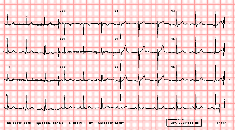
what if the history is concerning but ECG is normal?
An ECG showing ST depressions, TWI or STE is obviously
concerning. Patients who present with chest pain with suspected cardiac ischemia
based on the history but normal ECG should still undergo further diagnostic
testing with cardiac markers before they can be confidently assigned to a low
risk group.
What
if the history is concerning with ongoing ischemic symptoms, but ECG is normal
and troponin is not elevated?
This sounds very much like Unstable Angina. DO NOT SEND ANYONE
HOME WITH CONCERNING AND PERSISTING SYMPTOMS DESPITE NEGATIVE TROPONINS.
Unstable Angina can occur when you are resting, sleeping, or during little physical exertion. The pain may last longer than stable angina and rest or anti-ischemic medications usually do not help relieve it. USA can have an ischemic or normal ECG but should always have negative troponins by definition.
In contrast, Stable
Angina is very predictable with Chest Pain on exertion that gets better on
resting. Stable Angina us also relieved with anti-ischemic medications.
Beware
of the Non-Specific Troponinemia AKA Troponinitis!
Troponins are the preferred and recommended markers of myocardial
necrosis. Read more about troponins here. But the new generation hs troponins
are extremely sensitive and thus less specific i.e you might end up getting a
false positive elevated troponin leading to unnecessary admissions and work ups. So if history is not suggestive of ACS but
troponin is elevated – get a few more ECGs but do think of other possible
causes of an elevated troponin such as:


ACS
includes STEMI, NSTEMI, USA (not stable angina)
Patients with STEMI do not require troponin since their initial
treatment is determined by their clinical presentation and ECG findings. Patients
with STEMI are identified quickly, assigned a high risk category and have a
well-defined treatment strategy (ie. urgent reperfusion with PCI or
thrombolytics).


When does NSTEMI need immediate cathlab:
The ACC/AHA guidelines for NSTEMI recommend < 2 hour cath for:
- Refractory ischemia
- Ischemia with hemodynamic or electrical instability
If you are worried about a patient, get serial ECGs, send
troponins and involve cardiology at the earliest.
Disposition of a Low Risk Patient – Slightly concerning history but non-ischemic
ECG and negative enzymes.
Here we are specifically talking about Unstable Anginas which can be further divided into two groups i.e negative troponin with ischemic ECG and negative troponin with a non-ischemic ECG.
Current data
shows that if patients have negative troponins with non-ischemic ECG, then prognosis is not bad
even if they are diagnosed with unstable angina. If they have
unstable angina with an ischemic EKG, then they have a worse prognosis.
Note - if you see an Ischemic ECG – Get worried even when if the
enzymes are normal
Low risk unstable angina with negative troponins can have:
Shared Medical
Decision Making - Do serial troponins and serial ECGs. Current evidence suggests a repeat troponin at hour 3 from initial
EKG reduces potential miss rate from 1.7% to <1% at 30 days. Let them make this decision - ask them if they would want to get admitted or if they are happy to follow up as an out-patient with a week.


Use Clinical Decision Making rules such as HEART/GRACE score to further risk stratify them and most important - Document medical decision-making and Clinical Decision rules in the patient's record.
Read more on HEART SCORE on REBELEM.
Take Home:
- A good history helps in risk stratification. Don’t rule out ACS just based on the history alone. With the slightest of concern, get an ECG.
- It is okay to send troponins on your patients if you have some degree of concern but If there are no concerns eat all, then do not send troponins.
- Patients who present with chest pain with suspected cardiac ischaemia based on the history but normal ECG should still undergo further diagnostic testing.
- USA can have an ischemic or normal ECG but should always have negative troponins by definition.
- Low Risk - Do Serial ECGs, Shared Decision Making, Clinical Decision Making Rules to further risk stratify and DOCUMENT the decision making in the medical record.
Further Reading:
- http://hqmeded-ecg.blogspot.co.uk/2014/04/unstable-angina-dr-braunwald-asks-if-it.html
- http://hqmeded-ecg.blogspot.co.uk/2015/06/unstable-angina-still-exists-beware.html
- https://blog.essentialsofem.com/2016/02/25/low-risk-chest-pain-adp-showdown-using-timi-vs-heart-pt-1-of-3-timi/
Author:

Lakshay Chanana
Speciality Doctor
Northwick Park Hospital
Department of Emergency Medicine
England
No comments:
Post a Comment