Showing posts with label Cardiology. Show all posts

Monday, August 6, 2018

Pulmonary Hypertension

Introduction
Pulmonary vascular system is a high-flow, low-resistance circuit. Normal pulmonary arterial systolic pressures range from 15 to 30 mm Hg, whereas diastolic pulmonary arterial pressures range from 4 to 12 mm Hg and Pulmonary hypertension is defined as a mean pulmonary arterial pressure >25 mm Hg at rest or >30 mm Hg during exertion.

Although echocardiography can estimate pulmonary arterial pressure in a patient with suspected pulmonary hypertension, definitive diagnosis requires right heart catheterization. The World Health Organization classifies pulmonary hypertension into five categories based on cause and response to treatment:


Accurate classification of pulmonary hypertension is key to directing treatments. Regardless of the cause, patients with pulmonary hypertension have high morbidity and mortality rates.

Pathophysiology
Endothelial dysfunction results in an imbalance between endogenous vasodilators and vasoconstrictors with net effect leading to vasoconstriction and formation of in situ thrombi. Other pathologic processes include alterations in microvascular permeability, abnormal hypoxic vasoconstriction, microvascular thrombosis, and the formation of plexiform lesions, leading to vascular remodeling. 

Ultimately, these abnormalities result in sustained elevations of pulmonary vascular resistance and impairment of pulmonary blood flow leading to RV dilatation and poor contractility. With progressive RV dilation, the intraventricular septum is displaced toward the left ventricle. This displacement inhibits left ventricular filling and ultimately impairs cardiac output and systemic perfusion.

Clinical Presentation 

  • Symptoms can be non-specific which often leads to delayed diagnosis
  • Dyspnea, fatigue, chest pain, near syncope, syncope, exertional lightheadedness
  • Orthopnea, paroxysmal nocturnal dyspnea, and peripheral edema
The physical examination is often normal in the early stages of pulmonary hypertension.

Late signs include a holosystolic tricuspid regurgitation murmur, jugular venous distention, hepatomegaly, ascites, and lower extremity edema

Diagnosis 
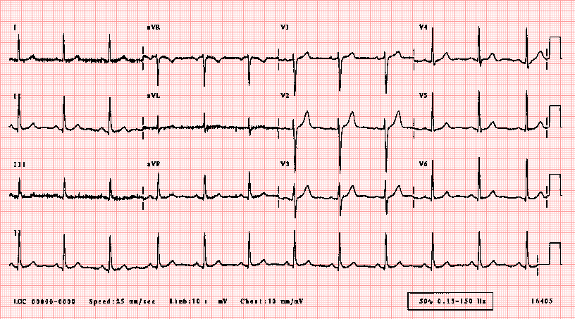
ECG: The most common ECG abnormality seen in pulmonary hypertension patients is right axis deviation. Additional findings associated with pulmonary hypertension include an R/S ratio >1 in lead V1, an R/S ratio<1 in leads V5 and V6, a qR complex in lead V1, an S1Q3T3, right atrial enlargement in the inferior leads, and an incomplete or complete right bundle branch block. The most common dysrhythmias in patients with pulmonary hypertension are atrial fibrillation, atrial flutter, and atrioventricular nodal reentrant tachycardia.


B-type natriuretic peptide and N-terminal B-type natriuretic peptide are often elevated  Elevations in troponin from myocardial ischemia or a strain-induced leak can be seen.

CXR: Common abnormalities associated with pulmonary hypertension include enlargement of the right atrium, RV, and hilar pulmonary arteries. 

TTE: Transthoracic echocardiography is the best initial diagnostic test to assess pulmonary hypertension in the ED. It allows estimation of the pulmonary artery systolic pressure and detection of decreased RV function, right atrial hypertrophy right ventricular hypertrophy and leftward deviation of the intraventricular septum.

page455image36080page455image36240page455image36400


Treatment 
No consensus guidelines exist for the management of critically ill patients with pulmonary hypertension in the ED. The mainstays of ED therapy include:

1. Supplemental oxygen (Target SpO2 >90%)
2. Optimizing intravascular volume, augmenting right ventricular function, maintaining coronary artery perfusion, and decreasing right ventricular afterload 

Treatments caveats:
  • Intubation: In patients with severe pulmonary hypertension, intubation and venti- lation can cause rapid cardiovascular collapse due to increased intrathoracic pressure from positive-pressure ventilation and effects of sedative medications on right ventricular function and systemic vascular resistance. Adjust the respiratory rate to avoid hypercapnia, which can increase pulmonary vascular resistance, pulmonary artery pres- sure, and RV strain.
  • Fluids in RV Failure: Volume overload can cause RV dilation, displacing the intraventricular septum, impairing left ventricular output, and ultimately compromising tissue perfusion.3 For patients who are hypovolemic, give serial boluses of an isotonic crystalloid solution in 250- to 500-mL aliquots with close monitoring.
  • RV Dysfunction: Dobutamine is preferred inotrope of choice. Avoid doses>10 micrograms/kg/min, because large doses can increase pulmonary vascular resistance and cause tachydysrhythmias and hypotension. For patients unable to tolerate dobutamine, milrinone is an alternative. Higher doses of milrinone can cause hypotension.
  • RCA Perfusion: For the hypotensive pulmonary hypertension patient, use a vasopressor to increase aortic root pressure and maintain RCA perfusion. Norepinephrine is recommended for this purpose. Avoid high doses of nor- epinephrine because it can increase pulmonary vascular resistance and impair right ventricular output.
  • RV Afterload reduction: These medications are rarely used in ED setting. Reducing right ventricular afterload with pulmonary vasodilators is a critical component in the management of stable patients with pulmo- nary hypertension. The most commonly used pulmonary vasodilators are prostanoids, endothelin receptor antagonists, and phosphodiesterase-5 (PDE-5) inhibitors. These medications are used primarily in the treatment of patients with pulmonary arterial hypertension. 

Prostanoids (epoprostenol, treprostinil, and iloprost) are potent vasodilators and are the initial treatment of choice in patients with pulmonary arterial hypertension and right ventricular failure. These medications have antiplatelet and antiproliferative properties.

Endothelin receptor antagonists (Currently not used for critically ill ED patients) are administered orally and increase exercise capacity, improve hemodynamics, and can delay the time to clinical worsening in pulmonary hypertension patients. Drugs: bosentan and ambrisentan. 

PDE-5 inhibitors (Currently not used for critically ill ED patients) sildenafil and tadalafil are approved for use in patients with pulmonary hypertension. They are administered orally, seeking to improve hemodynamics and exercise capacity in patients with pulmonary arterial hypertension.

Pulmonary HTN patients presenting to ED are often critically ill and require HDU/ICU level care. Therefore, almost all of them require admission with expert input. 


Posted by:

              
     Lakshay Chanana
     
     ST4 Trainee
     Royal Infirmary of Edinburgh
     Department of Emergency Medicine
     Edinburgh
     Scotland

     @EMDidactic


Monday, May 14, 2018

The Gastrocardiac Syndrome

Roemheld syndrome (RS) or gastric-cardiac syndrome, is a complex of gastrocardiac symptoms where issues with gut are found to be associated with cardiac symptoms like arrhythmias and benign palpitations. Despite a full workup, when the cause of symptoms is not found then this condition is one of the possible diagnosis. These are the patients who are often labeled as  "Non-Cardiac Chest Pain' or "MSK related CP" in the Emergency Department. 

Possible reasons for Gastrocardiac Syndrome
  • Anatomical close proximity between the stomach and the heart (GERD, Hiatus Hernia)
  • Same nerve supply - Vagus Nerve (Compressed by distended stomach leaading to bradycardia and occasionally arrythmias)
  • PPIs (Often used for GERD/Peptic ulcer) leading to HypoMg leading to ectopic beats

Due to pressure in the epigastric and left hypochondrium, the diaphragm is elevated and displaces the heart. This reduces the heart's ability to fill and increases the contractility of the heart causing palpitations/dizziness/anxiety. Typically, there are no changes/abnormalities related in the EKG detected. 


Symptoms can be variable and include a whole list of GI and Cardiovascular symptoms:  They are usually seen after eating a meal.
  • Sinus Bradycardia
  • Shortness of Breath
  • Chest tightness, Anxiety
  • Muscle crampiness
  • Dizziness
  • Palpitations
  • Belching, nausea

Conditions that come under the spectrum of RS:
  • GERD
  • Hiatus Hernia
  • Lactose intolerance
  • Gall Bladder disease
  • Intestinal Disorders 
RS in an ED diagnosis and our workup must include ECGs and troponins. Extensive GI and Cardiac workup are needed before calling this condition. Therefore, the diagnosis is made based on symptoms in the absence of heart abnormalities. 

Management
Ruling out another diagnosis such as peptic ulcer, bowel cancer, GB disease, ACS is the priority. Treatment is based on symptoms relief. Medications that have found to help include Anticholinergics, Antacids, Beta-Blockers, Anti-convulsants and simethicone. 

Take Home:
Gastrocardiac Syndrome is not an ED diagnosis. Always think of ACS first but beware of close proximity between stomach and heart.

References:

  1. Gastrocardiac syndrome: A forgotten entity; Saeed, Mohammad et al. The American Journal of Emergency Medicine , Volume 0 , Issue 0
  2. Jervell, O. and Lødøen, O. (1952), THE GASTROCARDIAC SYNDROME. Acta Medica Scandinavica, 142: 595-599. doi:10.1111/j.0954-6820.1952.tb13409.x
  3. The effect of the lateral decubitus position on vagal tone. Chen GY, Kuo CD. Anesthesia 1997 Jul;52(7):653-7. Department of Medicine, Provincial Tao-Yuan General Hospital, Republic of China.


Posted by:

              
     Lakshay Chanana
     
     Speciality Doctor
     Northwick Park Hospital
     Department of Emergency Medicine
     England

     @EMDidactic

Monday, July 3, 2017

When to stay away from Amiodarone

This week, listen to Amal Mattu talking about Amiodarone. 

Does Amiodarone really fix everything?


Monday, November 7, 2016

Chasing Chest Pains - Risk Stratification and Non Specific Troponinemia

Chest pain is caused by a myriad of causes ranging from benign to life threatening, some of which can cause death within minutes or hours. While evaluating chest pain, ACS is always high up on our list of differential diagnosis and as Emergency Physicians, it is our responsibility to not only robustly identify ACS and but also avoid needless investigations and unnecessary admissions for those who can be safely discharged from the ED after risk stratification.


Here is an overview of ACS with breakdown of terminologies, key points about history/physical exam and biomarkers and specifics about what to do with a low risk ACS patient:

Classical ACS presents with:
  •             Heavy, aching or tight
  •             Central chest or left sided
  •             Not related to respiration or movement
  •             May radiate to arms, neck, or jaw


You will often see patients who have one or a few of these features but end up having a completely negative work up for ACS. Remember, the history is helpful only to risk stratify – not to confirm your diagnosis. Everyone perceives pain in a different way but history is the first step during evaluation and risk stratification. 

Atypical ACS
Atypical presentations of ACS are common, occurring in up to 1/3rd of patients, mostly in the elderly, diabetics and women. Advanced age, co-morbid factors, delay in diagnosis lead to the increased mortality in these populations.


Things you must ask/look for:

  • Radiation to both arms – Likely  ACS
  • Radiation to left arm – Likely ACS
  • Nausea / Vomiting – Likely ACS
  • SOB on exertion – Likely ACS
  • Associated with Sweating – Likely ACS
  • Hypotension – Likely ACS
  • S3 – Likely ACS
  • Describes as previous angina - Likely ACS


  • Pleuritic/ Positional/ Sharp Pain – Unlikely ACS (not impossible)
  • Tender on Palpation – Unlikely ACS (not impossible)


A good history helps in risk stratification. Don’t rule out ACS just based on the history alone. With the slightest of concern, get an ECG.


Risk Factors
Risk factors once again help us to risk stratify but just based on the absence of risk factors you cannot rule out ACS. Get worried if the history is concerning even if there are no risk factors at all. The next step is ECG.


And what if the history is concerning but ECG is normal?
An ECG showing ST depressions, TWI or STE is obviously concerning. Patients who present with chest pain with suspected cardiac ischemia based on the history but normal ECG should still undergo further diagnostic testing with cardiac markers before they can be confidently assigned to a low risk group.


What if the history is concerning with ongoing ischemic symptoms, but ECG is normal and troponin is not elevated?
This sounds very much like Unstable Angina. DO NOT SEND ANYONE HOME WITH CONCERNING AND PERSISTING SYMPTOMS DESPITE NEGATIVE TROPONINS.

Unstable Angina can occur when you are resting, sleeping, or during  little physical exertion. The pain may last longer than stable angina and rest or anti-ischemic medications usually do not help relieve it. USA can have an ischemic or normal ECG but should always have negative troponins by definition.

In contrast, Stable Angina is very predictable with Chest Pain on exertion that gets better on resting. Stable Angina us also relieved with anti-ischemic medications.


Beware of the Non-Specific Troponinemia AKA Troponinitis!

Troponins are the preferred and recommended markers of myocardial necrosis. Read more about troponins here. But the new generation hs troponins are extremely sensitive and thus less specific i.e you might end up getting a false positive elevated troponin leading to unnecessary admissions and work ups.  So if history is not suggestive of ACS but troponin is elevated – get a few more ECGs but do think of other possible causes of an elevated troponin such as:


With hs Troponins, we are now able to pick up even the minimal level of troponins in a patient’s system and thus those who were diagnosed as Unstable Anginas (Ischemic Chest Pain/ECG but normal troponin levels) a decade back are now labeled as NSTEMIs (Ischemic Chest Pain/ECG but a raised troponin). At this moment, although it is controversial but some experts do believe that Unstable Angina does not even exist anymore in this era of highly sensitive troponins.


ACS includes STEMI, NSTEMI, USA (not stable angina)
Patients with STEMI do not require troponin since their initial treatment is determined by their clinical presentation and ECG findings. Patients with STEMI are identified quickly, assigned a high risk category and have a well-defined treatment strategy (ie. urgent reperfusion with PCI or thrombolytics).


When does NSTEMI need immediate cathlab:
The ACC/AHA guidelines for NSTEMI recommend < 2 hour cath for:
  1. Refractory ischemia
  2. Ischemia with hemodynamic or electrical instability


If you are worried about a patient, get serial ECGs, send troponins and involve cardiology at the earliest.


Disposition of a Low Risk Patient – Slightly concerning history but non-ischemic ECG and negative enzymes. 

Here we are specifically talking about Unstable Anginas which can be further divided into two groups i.e negative troponin with ischemic ECG and negative troponin with a non-ischemic ECG. 

Current data shows that if patients have negative troponins with non-ischemic ECG, then prognosis is not bad even if they are diagnosed with unstable angina.  If they have unstable angina with an ischemic EKG, then they have a worse prognosis.

Note - if you see an Ischemic ECG – Get worried even when if the enzymes are normal

Low risk unstable angina with negative troponins can have:
Shared Medical Decision Making -  Do serial troponins and serial ECGs. Current evidence suggests a repeat troponin at hour 3 from initial EKG reduces potential miss rate from 1.7% to <1% at 30 days. Let them make this decision - ask them if they would want to get admitted or if they are happy to follow up as an out-patient with a week. 


Use Clinical Decision Making rules such as HEART/GRACE score to further risk stratify them and most important - Document medical decision-making and Clinical Decision rules  in the patient's record.


Read more on HEART SCORE on REBELEM.


Take Home:
  • A good history helps in risk stratification. Don’t rule out ACS just based on the history alone. With the slightest of concern, get an ECG.
  • It is okay to send troponins on your patients if you have some degree of concern but If there are no concerns eat all, then do not send troponins.
  • Patients who present with chest pain with suspected cardiac ischaemia based on the history but normal ECG should still undergo further diagnostic testing.
  • USA can have an ischemic or normal ECG but should always have negative troponins by definition.
  • Low Risk - Do Serial ECGs, Shared Decision Making, Clinical Decision Making Rules to further risk stratify and DOCUMENT the decision making in the medical record. 


Further Reading:
  • http://hqmeded-ecg.blogspot.co.uk/2014/04/unstable-angina-dr-braunwald-asks-if-it.html
  • http://hqmeded-ecg.blogspot.co.uk/2015/06/unstable-angina-still-exists-beware.html
  • https://blog.essentialsofem.com/2016/02/25/low-risk-chest-pain-adp-showdown-using-timi-vs-heart-pt-1-of-3-timi/


Author:

              
     Lakshay Chanana
     
     Speciality Doctor
     Northwick Park Hospital
     Department of Emergency Medicine
     England

     @EMDidactic


Monday, October 31, 2016

Is troponin really becoming a lousy test?

Just a quick reminder this week regarding the use of troponin. This is one of the most abused tests in Emergency Departments these days.  The hs Troponins seems to be overtly sensitive that might lead to unnecessary admissions. Here are a few caveats with troponin:

1. Do a history first, followed by a physical exam and then ECG. Only after that, send a troponin if you are concerned about the possibility of ACS. 

DO NOT ORDER TROPONIN FOR EVERYONE WALKING IN WITH CHEST PAIN

2. Remember that there a  plenty of other reasons to have a positive troponin besides ACS. To name a few - PE, Heart Failure, Sepsis, Renal Failure, Stroke, Tachycardia, Infiltrative Heart Disease, Myopericarditis.

A RAISED TROPONIN DOES NOT ALWAYS EQUAL ACS. TROPONIN IS A MARKER OF ANY MYOCARDIAL INJURY, NOT JUST ISCHEMIA. 

3. STEMI does not need a troponin. Focus on rapid Reperfusion (Thrombolytics or PCI). Troponin is done to differentiate between NSTEMI and USA (Unstable Angina).


4. Discharging someone with a single negative troponin is high risk business (unless it was drawn 6 hours after the onset of symptoms). Whenever possible, get two serial troponins to see if there is any significant rise form the baseline. However, serial sampling of hs troponin at 0 and 2 hours can safely rule-out of STEMI and NSTEMI. 

In case, you happen to discharge home a patient with two negative troponins, document why are you doing that because it can still be Unstable Angina. 

5. Troponin might take 6-12 hours to rise from the onset of symptoms and stays elevated upto 2 weeks. Document your concerns while sending these cardiac biomarkers.


Attorneys focus on the documentation of HPI, documentation of ECG findings and Medical Decision Making with appropriate reasoning before settling the case. We are going to cover these bits next week. 


BOTTOMLINE: I think troponin is still a great test, if done with caveats. However, over the last two decades, it has become extremely sensitive and thus non-specific (like d-dimer for PE). 


Further Reading: 


Author:

              
     Lakshay Chanana
     
     Speciality Doctor
     Northwick Park Hospital
     Department of Emergency Medicine
     England

     @EMDidactic


Monday, August 1, 2016

Cardiogenic Pulmonary Edema: Hold on before pushing Diuretics!!

Case

A 65 year old female came to ER with sudden onset breathlessness. 

HR: 110/min
BP 180/90
RR 30/min
SpO2 90% on room air. 

Examination was characterised by bilateral coarse crepitations. She was known case of ischemic heart disease and hypertension, not compliant with her medications. 

The first differential that was unanimously thought was acute heart failure resulting in pulmonary edema. ECG showed sinus tachycardia, Chest X-ray confirmed diffuse pulmonary edema.

These cases form the bread and butter of Emergency Physician. Treating them wisely and quickly with appropriate drugs gives immediate relief. Many different drugs have been given as an early intervention - ranging from morphine, furosemide and nitroglycerin. But what actually works?


THE BUCKET THEORY by Amal Mattu
                   
Imagine the lung being the bucket with the tap as pre-load and hose (a flexible tube) as after-load. Increase in pre-load causes more water flowing in through faucet, thus filling the bucket. Similarly, an increase in afterload by reduced water out through the hose will lead to increased fluid in the bucket i.e. pulmonary edema. The third possibility being left ventricular dysfunction i.e. defects in the pump.


www.freeemergencytalks.net/2010/04/amal-mattu-winning-at-failure/

Causes of cardiogenic pulmonary edema  
  1. Excessive venous return (i.e increased preload) i.e. Tap is left open
  2. Excessive systemic vascular resistance (i.e. increased afterload) i.e. Hose is narrow
  3. LV dysfunction: a disorder of contractility or a disorder of rate and rhythm i.e. Pump failure


 Pathophysiology for cardiogenic pulmonary edema- a self-perpetuating cycle


GOALS OF TREATMENT 

     1. Turning the faucet (tap) off: Decreasing Preload

Traditional treatment: Morphine, Furosemide and Nitroglycerin

Morphine:
Action: Histamine release cause decrease in preload; anxiolysis may decrease catecholamine reducing afterload (As per studies by Vismara et al Circulation,1976)
Side-effects: Increase catecholamines: rash/urticarial; respiratory depression in high doses; direct myocardial depression

Mythbuster:
Timmis et al (BMJ 1980), gave 0.2mg/kg morphine to acute myocardial infarction patients with severe left ventricular failure found that 15min and 45 mins later, the heart rate, BP  and cardiac index reduced and there was no decrease in preload.

Hoffman et al (Chest 1987), morphine given in prehospital pulmonary edema patients had both subjective and objective deterioration.

From Acute decompensated heart failure national registry (ADHERE), Peacock WF et al (Emerg Med J 2008); compared morphine versus no morphine in acute decompensated heart failure. They observed that morphine administration increased the need for mechanical ventilation, ICU admission and prolonged hospital stay. Morphine was an independent predictor of mortality (OR: 4.84)

VERDICT: Morphine has NO ROLE in modern management of cardiogenic pulmonary edema

Furosemide

Action: Diuresis and venodilation
Increased afterload decreases renal blood flow, hence the action of furosemide is delayed by 30-120minutes in cardiogenic edema patients. The most common myth is doubling the dose of furosemide if the patient does not diurese, as a result after couple of hours the patient ends up becoming hypotensive. Diuretics work only when kidneys are perfused by reducing afterload.

Mythbuster:
Kiely et al (Circulation 1973), 15 post MI CHF patients, showed no drop in preload reduction due to veno-dilation, the drop occurred post diuresis.

Ikram et al (Clin Sci, 1980) showed a drop in cardiac output in 17% in first 90 minutes after furosemide administration. The cardiac output returned to baseline only after diuretic effect started.

Nelson et al (European Heart Journal 1983) showed a similar finding. The found a drop in cardiac output and stroke volume in first 30-60 minutes which returned to baseline after 60-90minutes.

Kraus et al Chest 1990, treated patients with furosemide, furosemide with nitrates and nitrates plus captopril before giving furosemide. If pre-medicated with nitrates and captopril, furosemide had an immediate and sustained effect on pulmonary capillary wedge pressure.

VERDICT: Diuretic effect is delayed if administered alone and can be detrimental in first 30-60 minutes. Furosemide activates the renin angiotensin aldosterone system and increases the afterload, causing more harm than good in pulmonary edema patients. So, when the patients undergo preload and afterload reduction, furosemide works immediately as the kidneys are perfused.

Nitroglycerin

Action: Immediate drop in preload and afterload
Advantages: Moderate to high doses bring significant reduction in systemic vascular resistance. It has a short half-life, can be titrated easily. Multiple modes of administration- sublingual, oral, intravenous and topical.
Side-effect: Head ache

Take caution if the patient is hypotensive, acute mitral regurgitation, aortic stenosis, pulmonary hypertension and patients on sildenafil

Being Aggressive! Starting at 50-60 microgram/min (NOT at 2.5-5mcg/min) and escalating 10-20mcg/min (upto 400mcg/min). Another way of optimising hemodynamics is thru a NTG bolus of 0.5 - 2mg. Multiple studies like Cotter et al 1998, Beltrame et al 1998, Kruas et al 1990, Hoffman et al 1987, Nelson et al 1983 have proved the importance of high dose nitroglycerin over smaller dosages.

The oral dose of NTG is 0.4mg or 400mcg is approximately equal to 80mcg/min infusion. Therefore, starting an infusion at 5mcg/min after administering 0.4mg sublingual dose leads to a dramatic dose reduction. 

VERDICT: Nitroglycerin should be the FIRST line drug for treatment of moderate CHF and pulmonary edema.


2. Using a bigger/ wider hose: Decreasing afterload

Nitroglycerin, nitroprusside, hydralazine and ACE-I

Nitroprusside can be used in acute mitral regurgitation and severe hypertension. But it has shown to have high fluctuations in BP and is not easily titrable. Similarly hydralazine is difficult to titrate.

ACE-I like captopril 25mg dipped in water placed sublingually for BP>110, has shown to be an excellent pre-load reducer in 10 minutes (Barnett et al, Curr Ther Research 1991), without any effect on heart rate and mean arterial pressure.

Langes et al (Current Ther Research 1993), showed that giving captopril infusion in moderate CHF with pulmonary edema decreases preload and afterload in 6 minutes, improves cardiac output without any adverse effects.

Varriale et al (Clin Cardiology 1993), showed that intra venous enalaprilat in severe CHF and mitral regurgitation, increased cardiac output and stroke volume, decreased preload and afterload, also decreased the magnitude of mitral regurgitation.

Saccheti et al (Am J Em Med 1999), showed that sublingual captopril significantly dropped the need for intubation or ICU admission in severe CHF patients (0.28:1).

Non-compliant dialysis patients who developed pulmonary edema showed improved outcome with sublingual captopril. (Saccheti et al Am J Em Med 1993).

VERDICT: Sublingual or intravenous ACE-I showed hemodynamic and subjective improvement in 6-12 minutes. Combination with IV nitroglycerin exceeds benefit with either drug alone. It can be used as an acceptable alternative to IV nitroglycerin.

Non Invasive Ventilation
Action: decreases preload and afterload, maintains positive airway pressure during respiration by keeping the stiff alveoli open and promoting gas exchange.

VERDICT: NIV decreases work of breathing, improves O2/ CO2 exchange, reduces length of stay, reduces hospital costs, reduces need for intubation, and may decrease mortality. Early NIV usage is a must!


3. Improving pump function: using ionotropic support

Patients with STEMI, where pump function is affected, there arises a need for ionotropic support.

Catecholamines: dobutamine, dopamine, phosphodiesterase inhibitors : milrinone, IABP (bridging device before PTCA/CABG)
No good literature to establish superiority of one over another.

VERDICT: Use what you are comfortable with. Use milrinone in cardiogenic pulmonary edema patients unresponsive to dobutamine/ dopamine.

More than 50% of patients in cardiogenic pulmonary edema are euvolemic. Fluid is excess in the wrong bucket. Hence treatment should be focussed towards ‘fluid redistribution’ and not ‘fluid removal’. 

Suggested algorithm for managing heart failure based on BP on ED arrival as normotensive failure, hypotensive failure and hypertensive failure.


Sauna for CHF patients!
Tie et al (Circulation, 1995) studied thermal vasodilation using sauna in 34 CHF patients and found that 15 minutes of sauna reduced preload, afterload, mitral regurgitation and significant improvement in cardiac index, stroke volume and ejection fraction!


SUMMARY
  • Treatment of pulmonary edema should be focused on ‘fluid redistribution’ and not ‘fluid removal’. 
  • First line: Nitroglycerin and NIV (start ASAP)
  • Second line: ACE-I (in addition or instead of NTG)
  • Third line: Diuretic like furosemide  
  • Morphine has NO ROLE in modern management of cardiogenic pulmonary edema

Listen to Dr. Mattu's talk on Pulmonary Edema 


Further Reading 
  1. Beltrame JF, Zeitz CJ, Unger SA, et al. Nitrate therapy is an alternative to furosemide/morphine therapy in the management of acute cardiogenic pulmonary edema. J Card Fail (1998) 4:271-9.
  2. Biddle TL, Yu PN. Effect of furosemide on haemodynamic and lung water in acute pulmonary edema secondary to myocardial infarction. Am J Cardiol 1979;43:86-90.
  3. Buseman W, Schupp D. Effect of sublingual nitroglycerin in emergency treatment of severe pulmonary edema. Am J Cardiol 1978;41:931-6.
  4. Cotter G, Metzkor E, Kaluski E, et al. Randomized trial of high-dose isosorbide dinitrate plus low-dose furosemide versus high-dose furosemide plus low-dose isosorbide dinitrate in severe pulmonary oedema. Lancet 1998;351:389-93.
  5. Figueras J, Weil MH. Blood volume prior to and following treatment of acute cardiogenic pulmonary edema. Circulation 1978;57:349-55.
  6. Gammage M. Treatment of acute pulmonary oedema: diuresis or vasodilatation? Lancet 1998;351:382-3.
  7. Kraus PA, Lipman J, Becker PJ. Acute preload effects of furosemide. Chest 1990;98:124-8.
  8. Mattu A, Sharma S, Perkins AM, Zevitz ME: Pulmonary edema, cardiogenic. eMedicine Journal 2002;3(2)
  9. Mattu A. Pulmonary edema. Emergency Physicians Monthly 2002;9(9):1,4-8,12,16,22.
  10. Tei C, Horikiri Y, Park JC, et al. Acute hemodynamic improvement by thermal vasodilation in congestive heart failure. Circulation 1995;91:2582-90. 
  11. Barnett JC, Zink KM, Touchon RC. Sublingual captopril in the treatment of acute heart failure. Curr Ther Res 1991;49:274-81.
  12. Avoiding Common Errors in the Emergency Department: Mattu, Shanmugam, Swadron, Tibbleg, Woolridge
  13. Cardiovascular Emergencies : Amal Mattu
  14. Heart Failure, Clinical Pathways in Emergency Medicine: Prof. Suresh S David 

Author:


Nikhil N. Tambe - @nikhil16mar
M.B.B.S., ECFMG (USA)


Emergency Medicine Resident (PGY-2)
Masters in Emergency Medicine (GWU)
Kokilaben Dhirubhai Ambani Hospital, Mumbai
Instructor (American Heart Association)
Lifesupporters Institute of Health Sciences, Mumbai


I am an Emergency Medicine enthusiast, a lifelong learner, a proponent of Evidence Based Medicine, love ECGs, resuscitation and emergency cardiology. After my graduation I worked as a Research Assistant in the USA. I have always been actively involved in teaching basic and advanced lifesaving skills to medics, paramedics and lay public since 2012. Currently I am pursuing EM Residency at Kokilaben Dhirubhai Ambani Hospital under George Washington University - Masters in Emergency Medicine Program. My vision is to create awareness about EBM and develop EM as a stand alone speciality in India.