Monday, May 28, 2018

Nasal Fractures - ED Management

Nasal fracture is a common ED presentation. Very often, its management primarily involves only simple reassurance and arranging timely follow up. 


The nasal pyramid is formed by two rectangular-shaped bones that articulate with the frontal bone, the frontal process of the maxilla, and the perpendicular plate of the ethmoid. A large proportion of the structural integrity is maintained by a cartilaginous framework of the nasal septum, lateral processes, and medial and lateral crura of the alar cartilages.


Examination
Look for bony crepitus, deformity, and edema
Profuse epistaxis suggests nasal fracture. 
Nasal bone mobility is checked by grasping the dorsum of the nose between the thumb and index finger and attempting to rock the nasal pyramid back and forth.
Perform anterior rhinoscopy after applying topical vasoconstrictors and evacuation of clots 

Important examnation findings:

  • Septal Hematoma

Failure to identify and treat a septal hematoma can result in a saddle deformity of the septum, which will require surgical repair. A septal hematoma is a blood-filled cavity between the cartilage and the supporting perichondrium. If left untreated, these pockets of blood easily become infected. The resulting necrosis of the underlying cartilaginous support may result in per- manent saddle nose deformity
  • Mucosal lacerations
  • Head/C Spine trauma
  • Other facial bone injuries
  • Extraocular movements, VA
  • CSF leak
Management
Nasal bone fracture is a clinical diagnosis. Radiologic confirmation of isolated nasal fracture is not required as results of plain films rarely change management. Most nasal fractures do not require immediate intervention and are managed at ENT follow-up within 7-10 days. In ED, ruling out siginificant head trauma is the prioroty in addition to looking for a septal hematoma. Nasal fractures with overlying lacerations are treated as open fractures. Here is a flow chart suggesting ED managemnt of Nasal Bone Fracture:


During ED visits, soft tisssue edema obscures adequate physical examination. Thus, it is recommended to go for an ENT consultation for elective closed reduction in about 7-10 days. Delayed presnetations may develop fibrous connective tissue along the fracture line and lead to worse cosmetic outcome require rhinoseptoplasty. 


References:
  • https://www.ncbi.nlm.nih.gov/pmc/articles/PMC2579544/pdf/523.pdf
  • https://www.aafp.org/afp/2004/1001/p1315.pdf


Take Home:
ED Management of nasal fracture relies on ruling out other potential injuries (Head, C Spine, Face) and looking for local complications such as profuse bleeding and septal hematoma. Most injuries can be managed with reasuurance and pain relief and arranging an ENT follow up in a week. 


Posted by:

              
     Lakshay Chanana
     
     Speciality Doctor
     Northwick Park Hospital
     Department of Emergency Medicine
     England

     @EMDidactic


Monday, May 21, 2018

Bell's Palsy

Bell's palsy is an idiopathic LMN palsy involving the facial nerve accounting for up to 3/4 of all cases of unilateral facial paralysis. It is believed that inflammation and swelling of the facial nerve causes compression of the nerve which manifests as facial muscle weakness. However, the exact etiology is controversial. Reactivated herpes simplex virus is thought to be the most probable cause followed by herpes zoster virus.

The facial nerve consists of a:
  • Motor component, which supplies all the muscle of facial expression
  • Sensory branch that carries taste sensation from the anterior two thirds of the tongue through the chorda tympani nerve
  • Parasympathetic fibres reach the lacrimal glands via the greater superficial petrosal nerve, and they reach the sublingual and submaxillary glands via the chorda tympani. 
Bell's palsy is diagnosed upon the abrupt onset of unilateral facial weakness or complete paralysis of all the muscles on one side of the face, dry eye, pain around the ear, an altered sense of taste, hyperacusis, or decreased tearing. On attempted closure, the eye rolls upward (Bell's phenomenon). The disease usually progresses from the onset of symptoms to maximal weakness within three days. 

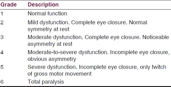
The most wodely used classification to grade severity of the facial muscle weakness is House-Brackmann classification:


UMN and LMN facial palsy

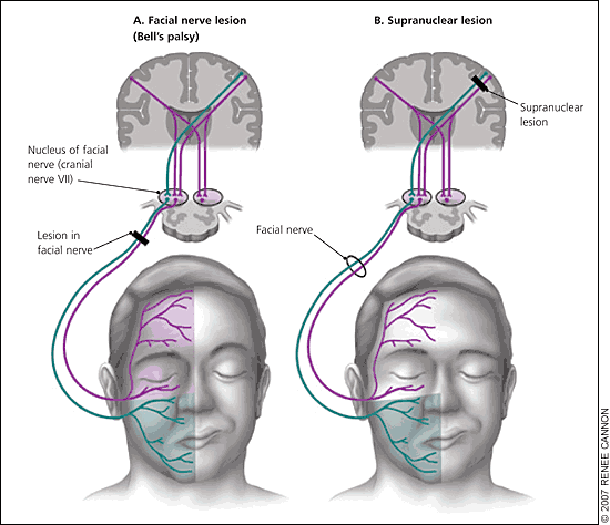
Classically stroke presents with a UMN type of facial nerve palsy i.e. a supra nuclear lesion sparing of forehead/eyelid muscles (image on right) and lesions beloew the facial nerve nuclei presnet with involvement of upper as well as lower half of face i.e involving forehead/eyelid muscles as well. It is important to look for involvement of limb weakness and deficts in other cranial nerves fucntion as LMN palsy may reperesent a brainstem stroke. 


Diagnosis and Treatment 


Focus on the history and physical examination to identify the possible causes of LMN facial palsy such as:

  • Otitis media
  • Trauma
  • Postsurgical complications
  • Neoplasms
  • Sarcoidosis
  • Lyme Disease 
  • Reactivation of VZV infection


Corticosteroids adminiatered within 3 days of the onset of synmptoms increase the likelihood of recovery and shorten time to recovery as well. Patients with Bell's palsy do not complain of any facial pain and don't have any cranial nerve involvement other than the facial nerve.

Recent literature favors the use of steroids and not antivirals. Sullivan et al in 2007 examined the treatment options for BP in a randomized control trial across 17 sites in Scotland. At three months, 83% of patients in the prednisolone group versus 63.6% of patients in the non-prednisolone group fully recovered. A meta-analysis in 2009 determined there was no significant benefit of combined antiviral and steroid treatment compared to steroids alone. Currently, the recommended treatment regiment for BP is prednisone, 60 to 80 mg per day, for one week or giving 25 mg twice daily for 10 days. 


Eye Care: Prescribe lubricating eye drops for use during the day in addition to a corneal lubricant to use at night. Furthermore, patches or taping the eyelid closed can be used at night. 


Severe disease can result in inability to completely close the eye in addition to decreased lacrimal secretions, leading to drying and tearing of the cornea. 

Summary

  • Bell’s palsy is idiopathic paralysis of the facial nerve and is the most common cause of LMN facial palsy. DO a full neuro exam and look for other cranila nerve involvement to look for potential brainstem CVA.
  • Bell's Palsy is unilateral and acute in onset, progressing over a period of hours and reaching maximal intensity within several days
  • Treatment consists of corticosteroids and eye care. The prognosis of BP is excellent, with 85% of patients regaining function within three weeks

References:
  1. Fahimi J, Navi BB, Kamel H. Potential misdiagnoses of Bell's palsy in the emergency department. Annals of emergency medicine. 2014 Apr 1;63(4):428-34.
  2. Sullivan FM, Swan IR, Donnan PT, Morrison JM, Smith BH, McKinstry B, Davenport RJ, Vale LD, Clarkson JE, Hammersley V, Hayavi S, McAteer A, Stewart K, Daly F. Early treatment with prednisolone or acyclovir in Bell’s palsy. N Engl J Med. 2007 Oct 18;357(16):1598-607.
  3. https://cks.nice.org.uk/bells-palsy#!scenario
  4. Quant EC, et al. The benefits of steroids versus steroids plus antivirals for treatment of Bell’s palsy: a meta-analysis. BMJ. 2009;339:b3354 
Posted by:

              
     Lakshay Chanana
     
     Speciality Doctor
     Northwick Park Hospital
     Department of Emergency Medicine
     England

     @EMDidactic

Monday, May 14, 2018

The Gastrocardiac Syndrome

Roemheld syndrome (RS) or gastric-cardiac syndrome, is a complex of gastrocardiac symptoms where issues with gut are found to be associated with cardiac symptoms like arrhythmias and benign palpitations. Despite a full workup, when the cause of symptoms is not found then this condition is one of the possible diagnosis. These are the patients who are often labeled as  "Non-Cardiac Chest Pain' or "MSK related CP" in the Emergency Department. 

Possible reasons for Gastrocardiac Syndrome
  • Anatomical close proximity between the stomach and the heart (GERD, Hiatus Hernia)
  • Same nerve supply - Vagus Nerve (Compressed by distended stomach leaading to bradycardia and occasionally arrythmias)
  • PPIs (Often used for GERD/Peptic ulcer) leading to HypoMg leading to ectopic beats

Due to pressure in the epigastric and left hypochondrium, the diaphragm is elevated and displaces the heart. This reduces the heart's ability to fill and increases the contractility of the heart causing palpitations/dizziness/anxiety. Typically, there are no changes/abnormalities related in the EKG detected. 


Symptoms can be variable and include a whole list of GI and Cardiovascular symptoms:  They are usually seen after eating a meal.
  • Sinus Bradycardia
  • Shortness of Breath
  • Chest tightness, Anxiety
  • Muscle crampiness
  • Dizziness
  • Palpitations
  • Belching, nausea

Conditions that come under the spectrum of RS:
  • GERD
  • Hiatus Hernia
  • Lactose intolerance
  • Gall Bladder disease
  • Intestinal Disorders 
RS in an ED diagnosis and our workup must include ECGs and troponins. Extensive GI and Cardiac workup are needed before calling this condition. Therefore, the diagnosis is made based on symptoms in the absence of heart abnormalities. 

Management
Ruling out another diagnosis such as peptic ulcer, bowel cancer, GB disease, ACS is the priority. Treatment is based on symptoms relief. Medications that have found to help include Anticholinergics, Antacids, Beta-Blockers, Anti-convulsants and simethicone. 

Take Home:
Gastrocardiac Syndrome is not an ED diagnosis. Always think of ACS first but beware of close proximity between stomach and heart.

References:

  1. Gastrocardiac syndrome: A forgotten entity; Saeed, Mohammad et al. The American Journal of Emergency Medicine , Volume 0 , Issue 0
  2. Jervell, O. and Lødøen, O. (1952), THE GASTROCARDIAC SYNDROME. Acta Medica Scandinavica, 142: 595-599. doi:10.1111/j.0954-6820.1952.tb13409.x
  3. The effect of the lateral decubitus position on vagal tone. Chen GY, Kuo CD. Anesthesia 1997 Jul;52(7):653-7. Department of Medicine, Provincial Tao-Yuan General Hospital, Republic of China.


Posted by:

              
     Lakshay Chanana
     
     Speciality Doctor
     Northwick Park Hospital
     Department of Emergency Medicine
     England

     @EMDidactic

Monday, May 7, 2018

GABHS Pharyngitis - Strep throat

Group A Î²-hemolytic Streptococcus pharyngitis is the most commonly occurring form of acute bacterial pharyngitis primarily affecting children age 5-15 years old. It is typically a self-limited disease where symptoms diminish markedly at days 3 and 4 after symptom onset, and antibiotics only decrease the duration of symptoms by approximately 16 hours. Use of antibiotics for everyone with strep throat is controversial with but most physicians are not comfortable withholding antibiotics. Read this post on REBEL EM questioning the use Abx in strep throat. 

Symptoms of acute pharyngitis:
  • Sore throat, cough, hoarseness, coryza
  • Odynophagia
  • Fever
  • Headache, abdominal pain, nausea and vomiting, diarrhea
  • Arthralgias, myalgias,lethargy

Symptoms suggestive of viral infection:
  • Conjunctivitis, Coryza, Cough
  • Diarrhea, Hoarseness, Viral Exanthem
Physical examination findings:
  • Tonsillopharyngeal erythema and/or exudates
  • Soft palate petechiae, uvulitis
  • Anterior cervical lymphadenitis


It is difficult to distinguish between viral and bacterial causes based on physical examination alone, and tonsillar exudate does not imply bacterial etiology. Bacterial etiology is often overdiagnosed and unnecessary antibiotic treatment is given. 


Several clinical prediction rules have been created to identify cases of GABHS pharyngitis, and a commonly used rule is FeverPAIN score (recommended by NICE guidelines). 

Diagnosis

Performing diagnostic testing depends on local protocols but it is generally done by obtaining a swab from the throat and doing rapid antigen detection test (RADT) and/or culture because the clinical features alone do not reliably discriminate between GAS and viral pharyngitis. Routine use of backup throat cultures for those with a negative RADT is not necessary for adults. Anti-streptococcal antibody titers are not recommended in the routine diagnosis of acute pharyngitis as they reflect past but not current events. 
  • Gold standard for diagnosis - Bacterial culture (sensitivity of 90% to 95%)
  • Rapid antigen detection (highly specific, sensitivity 80% to 90%) 


Current American guidelines recommend confirmatory throat culture for all patients with a negative antigen test. The antibiotic treatment of GABHS pharyngitis shortens the duration of illness, prevents transmission, prevent suppurative complications (acute otitis media, acute sinusitis, and peritonsillar abscess), and prevent systemic illness such as rheumatic fever, rheumatic heart disease, and post-streptococcal glomerulonephritis. Antibiotics for the treatment of GABHS pharyngitis should be reserved for those patients with a positive antigen test or culture, or those meeting clinical criteria for diagnosis. 


Here is a summary of NICE guidelines:


Decision Rules 

FeverPAIN score 
Centor Criteria 


Treatment
  • Symptom relief with NSAIDs
  • Local Anethetics sprays 
  • Antibiotics (Her is another perspective on Abx use in Strep throat)
  • Steroids - Emerging evidence to achieve better symptoms relief 
Antibiotic treatment can be delayed safely for a few days after symptom onset and still prevent major nonsuppurative sequelae. This provides us time to get confirmatory cultures. Also, there is no definitive evidence that antibiotic use can prevent acute glomerulonephritis. 

Penicillin remains the treatment of choice. A course of 10 days of oral therapy with twice-a-day dosing is recommended for complete pharyngeal eradication; similar efficacy is achieved with once-daily dosing of amoxicillin for 10 days. Clarithromycin and first-generation cephalosporins are alternatives in penicillin-allergic patients. Clindamycin may be required for macrolide-resistant GABHS in the penicillin-allergic patient. 


Take Home:
  • Know your local protocols about diagnostic testing and indications for antibiotics. 
  • Strep throat is typically a self-limited disease where symptoms diminish markedly at days 3 and 4
  • There is no definitive evidence that antibiotic use can prevent acute glomerulonephritis
  • It is difficult to distinguish between viral and bacterial causes based on physical examination alone.

References and Further Reading:
  1. Shaikh N, Swaminathan N, Hooper EG: Accuracy and precision of the signs and symp- toms of streptococcal pharyngitis in children: a systematic review. J Pediatr 160: 487, 2012. [PMID: 22048053]
  2. Bisno AL: Acute pharyngitis. N Engl J Med 344: 205, 2001. [PMID: 11172144]
  3. Centor RM, Witherspoon JM, Dalton HP, et al: The diagnosis of strep throat in adults in the emergency room. Med Decis Making 1: 239, 1981. [PMID: 6763125]
  4. Wong DM, Blumberg DA, Lowe LG: Guidelines for the use of antibiotics in acute upper respiratory tract infections. Am Fam Physician 74: 956, 2006. [PMID: 17002029]
  5. Shulman ST, Bisno AL, Clegg HW, et al: Clinical practice guideline for the diagnosis and management of group A streptococcal pharyngitis: 2012 update by the Infectious Diseases Society of America. Clin Infect Dis 55: e91, 2012. [PMID: 23091044]
  6. https://www.nice.org.uk/guidance/ng84/documents/draft-guidance
  7. https://academic.oup.com/cid/article/55/10/e86/321183
  8. https://www.nice.org.uk/guidance/ng84/chapter/summary-of-the-evidence#corticosteroids
  9. http://www.idsociety.org/uploadedFiles/IDSA/Guidelines-Patient_Care/PDF_Library/Strep.pdf

Posted by:

              
     Lakshay Chanana
     
     Speciality Doctor
     Northwick Park Hospital
     Department of Emergency Medicine
     England

     @EMDidactic