Showing posts with label Pulmonary Embolism. Show all posts

Monday, August 6, 2018

Pulmonary Hypertension

Introduction
Pulmonary vascular system is a high-flow, low-resistance circuit. Normal pulmonary arterial systolic pressures range from 15 to 30 mm Hg, whereas diastolic pulmonary arterial pressures range from 4 to 12 mm Hg and Pulmonary hypertension is defined as a mean pulmonary arterial pressure >25 mm Hg at rest or >30 mm Hg during exertion.

Although echocardiography can estimate pulmonary arterial pressure in a patient with suspected pulmonary hypertension, definitive diagnosis requires right heart catheterization. The World Health Organization classifies pulmonary hypertension into five categories based on cause and response to treatment:


Accurate classification of pulmonary hypertension is key to directing treatments. Regardless of the cause, patients with pulmonary hypertension have high morbidity and mortality rates.

Pathophysiology
Endothelial dysfunction results in an imbalance between endogenous vasodilators and vasoconstrictors with net effect leading to vasoconstriction and formation of in situ thrombi. Other pathologic processes include alterations in microvascular permeability, abnormal hypoxic vasoconstriction, microvascular thrombosis, and the formation of plexiform lesions, leading to vascular remodeling. 

Ultimately, these abnormalities result in sustained elevations of pulmonary vascular resistance and impairment of pulmonary blood flow leading to RV dilatation and poor contractility. With progressive RV dilation, the intraventricular septum is displaced toward the left ventricle. This displacement inhibits left ventricular filling and ultimately impairs cardiac output and systemic perfusion.

Clinical Presentation 

  • Symptoms can be non-specific which often leads to delayed diagnosis
  • Dyspnea, fatigue, chest pain, near syncope, syncope, exertional lightheadedness
  • Orthopnea, paroxysmal nocturnal dyspnea, and peripheral edema
The physical examination is often normal in the early stages of pulmonary hypertension.

Late signs include a holosystolic tricuspid regurgitation murmur, jugular venous distention, hepatomegaly, ascites, and lower extremity edema

Diagnosis 
ECG: The most common ECG abnormality seen in pulmonary hypertension patients is right axis deviation. Additional findings associated with pulmonary hypertension include an R/S ratio >1 in lead V1, an R/S ratio<1 in leads V5 and V6, a qR complex in lead V1, an S1Q3T3, right atrial enlargement in the inferior leads, and an incomplete or complete right bundle branch block. The most common dysrhythmias in patients with pulmonary hypertension are atrial fibrillation, atrial flutter, and atrioventricular nodal reentrant tachycardia.


B-type natriuretic peptide and N-terminal B-type natriuretic peptide are often elevated  Elevations in troponin from myocardial ischemia or a strain-induced leak can be seen.

CXR: Common abnormalities associated with pulmonary hypertension include enlargement of the right atrium, RV, and hilar pulmonary arteries. 

TTE: Transthoracic echocardiography is the best initial diagnostic test to assess pulmonary hypertension in the ED. It allows estimation of the pulmonary artery systolic pressure and detection of decreased RV function, right atrial hypertrophy right ventricular hypertrophy and leftward deviation of the intraventricular septum.

page455image36080page455image36240page455image36400


Treatment 
No consensus guidelines exist for the management of critically ill patients with pulmonary hypertension in the ED. The mainstays of ED therapy include:

1. Supplemental oxygen (Target SpO2 >90%)
2. Optimizing intravascular volume, augmenting right ventricular function, maintaining coronary artery perfusion, and decreasing right ventricular afterload 

Treatments caveats:
  • Intubation: In patients with severe pulmonary hypertension, intubation and venti- lation can cause rapid cardiovascular collapse due to increased intrathoracic pressure from positive-pressure ventilation and effects of sedative medications on right ventricular function and systemic vascular resistance. Adjust the respiratory rate to avoid hypercapnia, which can increase pulmonary vascular resistance, pulmonary artery pres- sure, and RV strain.
  • Fluids in RV Failure: Volume overload can cause RV dilation, displacing the intraventricular septum, impairing left ventricular output, and ultimately compromising tissue perfusion.3 For patients who are hypovolemic, give serial boluses of an isotonic crystalloid solution in 250- to 500-mL aliquots with close monitoring.
  • RV Dysfunction: Dobutamine is preferred inotrope of choice. Avoid doses>10 micrograms/kg/min, because large doses can increase pulmonary vascular resistance and cause tachydysrhythmias and hypotension. For patients unable to tolerate dobutamine, milrinone is an alternative. Higher doses of milrinone can cause hypotension.
  • RCA Perfusion: For the hypotensive pulmonary hypertension patient, use a vasopressor to increase aortic root pressure and maintain RCA perfusion. Norepinephrine is recommended for this purpose. Avoid high doses of nor- epinephrine because it can increase pulmonary vascular resistance and impair right ventricular output.
  • RV Afterload reduction: These medications are rarely used in ED setting. Reducing right ventricular afterload with pulmonary vasodilators is a critical component in the management of stable patients with pulmo- nary hypertension. The most commonly used pulmonary vasodilators are prostanoids, endothelin receptor antagonists, and phosphodiesterase-5 (PDE-5) inhibitors. These medications are used primarily in the treatment of patients with pulmonary arterial hypertension. 

Prostanoids (epoprostenol, treprostinil, and iloprost) are potent vasodilators and are the initial treatment of choice in patients with pulmonary arterial hypertension and right ventricular failure. These medications have antiplatelet and antiproliferative properties.

Endothelin receptor antagonists (Currently not used for critically ill ED patients) are administered orally and increase exercise capacity, improve hemodynamics, and can delay the time to clinical worsening in pulmonary hypertension patients. Drugs: bosentan and ambrisentan. 

PDE-5 inhibitors (Currently not used for critically ill ED patients) sildenafil and tadalafil are approved for use in patients with pulmonary hypertension. They are administered orally, seeking to improve hemodynamics and exercise capacity in patients with pulmonary arterial hypertension.

Pulmonary HTN patients presenting to ED are often critically ill and require HDU/ICU level care. Therefore, almost all of them require admission with expert input. 


Posted by:

              
     Lakshay Chanana
     
     ST4 Trainee
     Royal Infirmary of Edinburgh
     Department of Emergency Medicine
     Edinburgh
     Scotland

     @EMDidactic


Monday, May 22, 2017

Pulmonary Embolism Dilemmas

22/F on OCPs and recent history of prolonged air travel. She was found to have a DVT and was started on Rivaroxaban a week ago. She presented to the ED with c/o Sudden onset chest pain, pre-syncope and SOB. With a clot in her legs, her story was certainly concerning a PE. These were her Vitals on arrival:

PR 128/min, BP 110/70, RR 28/min, SPO2 99% on Room Air 
ECG - Sinus Tachy
PaO2 - 14kP (105mmHg)


I booked her for a CTPA on arrival since she was high-risk so d-dimer was not needed. Over the next hour, with some IV Fluids, her vitals settled down and looked a bit better.

PR 110/min BP 120/68 RR 24/min SPO2 99% Room Air

This made me wonder if a scan was really needed for her? Anyways, she was already on oral anticoagulation. Would I be able to justify myself for not doing a CTPA with textbook presentation of PE?

I ended up scanning her and she was found to have sub-segmental PEs. You might argue against a CTPA for this patient but her presentation and initial vitals kept me on my toes. 

My learning points from this case:

  • Normalization of initially abnormal vital signs should not be relied upon to r/o PE
  • ABG and pulse oximetry do not reliably predict the presence or absence of PE

When should we start a working up PE?
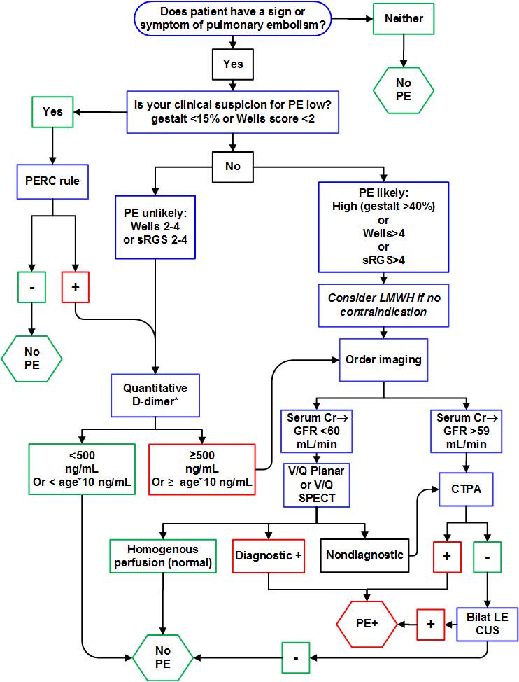
Start work up for PE if patient has any sign (Unexplained tachycardia/tachypnea, low SpO2) or symptom (Chest Pain, SOB) of PE. It is reasonable to consider PE as a differential when ECG shows evidence of right Heart Strain (S1Q3T3, Simultaneous TWI in inferior an pre-cordial leads). 

Caveat: All patients coming with Chest pain do not need a work up for PE. Risk Stratify them and score them on Well's criteria and PERC rule before starting the work up. Random ordering of d-dimer leads to false positive and unnecessary imaging/anticoagulation. 


Random d-dimer sent on a patient with Non-Specific Chest Pain. Can't we just ignore that?
D-Dimer is a great test if used sensibly. It is reassuring if negative (in low risk patients) to exclude PE. Always utilise d-dimer with clinical probability. 


If you are thinking high risk for PE, do not even bother sending a d-dimer. You will need imaging to rule out PE in high-risk regardless of the d-dimer results. As with every test in medicine, there are false positives and false negatives with d-dimer as well. We often talk about the non-specificity of d-dimer and false positives. Here is a list of things that cause false positive and false negative d-dimer.

Of course, you can disregard an elevated d-dimer but you should have a reasonable explanation for that. It should be documented clearly why imaging (CTPA or VQ) was not pursued if d-dimer was found elevated. 


When do we consider thrombolysis for PE?
PEs can be classified as:
  1. Massive PE is defined as acute PE with obstructive shock or SBP <90 mmHg
  2. Submassive PE is acute PE without systemic hypotension (SBP ≥90 mm Hg) but with either RV dysfunction or myocardial necrosis (positive troponin or ECHO evidence of RV dysfunction)
  3. Low risk PEs
Massive PE are definite ones for thrombolysis whereas thrombolysing Submissive PEs is a bit more controversial. Low risk PEs get anti coagulated with heparin. Other indications for using thrombolytics can be Cardiac Arrest with known PE patient or in someone with Presumed PE. 

Click here to read more about thrombolysis in PE. 


Troponin is not helpful in the diagnosis of PE, but it is helpful in the assessment for severity of disease.  


Do we need to treat small distal clots but without DVT (single clot <3mm)?
Isolated distal clots can represent a possible artefact rather than true disease. Many Sub segmental PEs are not even seen by another radiologist when blinded. But guidelines still recommend to treat if risk factors present or if symptomatic and has low risk of bleeding. 


What if CTPA turns negative but you have extremely high suspicion for PE?
Ask radiologist about the quality of scanner. Clinical context holds above everything else. Remember, Gold Standard for diagnosis is Pulmonary Angiography. 


When to consider IVC filter, cather directed lysis?
The indications for IVC filter include: 
(1) Patients with contraindications to anticoagulation
(2) Those who have complications from the use of anti-coagulation
(3) Those who fail to attain adequate anticoagulation while undergoing treatment. 


Cather directed lysis should be first line if available for massive PE. 


Working up PE in pregnancy? 
Start with lower extremity doppler (if DVT+ and hemodynamically stable, start anti-coagulation). Some authorities do not even recommend sending a d-dimer in pregnancy while others recommend using trimester adjusted d-dimer. When used in pregnancy, d-dimer can be extremely helpful when negative. Here is an algorithm based on using trimester adjusted d-dimer.


If you do not want to use d-dimer at all then you have these options:
  • Treat with Heparin Doppler if positive for DVT
  • ECHO/Troponin to further risk stratify
  • CTPA (Radiation is 4 times more than that of a V/Q scan)
  • VQ Scan (done only if CXR is normal, if CXR abnormal then go for CTPA)
             
                                         CTPA - Good for baby, Bad for Mom
                                         V/Q - Bad for baby, Good for Mom


Consider V/Q in cases of Allergy to iodine contrast, Impaired Renal Function and Pregnancy. Issues with V/Q scan can be availability, expertise to interpret the scan. V/Q often gets interpreted as low probability or intermediate probability which leaves you nowhere! 
Too many CTPAs can give you false positives and lead to over-diagnosis and over treatment. Ask yourself, if that tiny clot is really responsible for your patient's symptoms or was it just an incidetaloma! 

The least desirable scenario is one in which both a V/Q scan and a CTPA are needed to complete the diagnostic evaluation. 


Working up PE in a patient already on oral anticoagulants?
Anticoagulants can make d-dimer unreliable and there is no clear consensus on this. 
CTPA them if you have any concerns for a PE and plan for an IVC filter if they are CTPA positive. 


Consider targeting higher INR as they are throwing clots even on oral anti-coagulants. 


Summary:
The testing to catch Pulmonary Embolisms has skyrocketed over the last decade. The increased use of CT in patients with suspected PE has resulted in an increase in the diagnosis of PE but without an associated mortality benefit. Start a work up for PE based on your gestalt or Well's Criteria followed by PERC rule (Low risk on Well's Criteria and PERC negative does not even need a d-dimer). Every test comes with caveats (d-dimer, CTPA, VQ scan) and we must act in a prudent manner while ordering imaging to avoid unnecessary anti-coagulation. 


Further Reading
  • Age-Adjusted d-dimer
  • Tichauer M. The Emergency Medicine Approach To The Evaluation And Treatment Of Pulmonary Embolism. Emergency Medicine Practice. 2012:2.
  • Kline JA, Kabrhel C. Emergency evaluation for pulmonary embolism, Part 2: diagnostic approach. The Journal of emergency medicine. 2015 Jul 31;49(1):104-17.


Posted by:

              
     Lakshay Chanana
     
     Speciality Doctor
     Northwick Park Hospital
     Department of Emergency Medicine
     England

     @EMDidactic


Monday, December 26, 2016

Sorting out Gabrahat (Anxiety) ~ The Common Complaint in the ED

 Gabrahat (Anxiety) is often a common complaint with many implications.
While working in Emergency Departments in India I have been surprised with what the ultimate diagnosis was when I investigated Gabrahat.
For me Gabrahat is as vague as the Horizon and I take this complaint very seriously. It is very easy for any Nurse or Emergency Physician to get framed and just label Gabrahat as Anxiety or Hysteria.
This can be the Epic Blunder of Large Proportions. 

Many times relatives who accompany the patient will Frame the Emergency Physician by saying words like “There is Tension”. What they mean to imply is Gabarahat is Stress Related.

I often relate Gabrahat to a “SENSE OF IMPENDING DOOM”. When you grade GABRAHAT in that perspective, it guides the Emergency Physician to be very Proactive and diligent.


Let me share a few blasts from the past which I have modified for the sake of Education.


Case One:


Middle Age Female comes to the ED saying that she is feeling SOB. She is hyperventilating and Diaphoretic. She says that she has been having pain all over the body and fells GABRAHAT as if something is going to happen to her.
Her vitals are stable but she continues to breathe hard and breath fast. The relatives were doing a Fine job of Framing her as hysteria.

Rapid Fire Questionnaire Labs EKG Trop and a X-ray Beta HCG UA and a BNP are ordered.


On examining the patient the only Finding is the breathing. Lorazepam given IV and Oxygen started and ABG Ordered which is showing alkalosis. Aspirin given and a bedside Glucose is Normal. She settles down but continues to breathe hard. A CTA Chest is ordered. 
There are massive shower Pulmonary Emboli. Pt gets thrombolyzed and goes to ICU.

Case Two:


A 55-year-old women comes with GABRAHAT. She says that she is afraid something is going to happen. She has no other symptom. She has no Past Psyc Issues.
Labs EKG Trop and an X-ray UA ordered. She has had a prior hysterectomy.


She had an ST Elevation MI. Went to the Cath Lab. No Symptoms at all. No Past History at all.


Case Three:


30-Year-old man came saying He had Gabrahat and felt that there was Irritation in the Chest. NO PAIN BUT ONLY IRRITATION. Exam Past History negative.
Cardiac labs CBC RFT LFT was negative so was his EKG and Xray. Against the will of the Internal Medicine Colleagues Pt admitted. 4 hour repeat EKG and Trop was placed from the ED. His EKG was normal but his Trop had become positive.


Cardiology who scheduled the patient for a cath after admitting him to CCU found a Tight Lcx Lesion which needed a Stent.


Case Four:


48 Female with Gabrahat. Second visit after discharge from the hospital. Come back saying she is afraid. No Pain, No Focus of Infection. 
CBC RFT LFT Cardiac Labs X-ray Beta HCG and UA Negative.

Says her Mind tell her Something is wrong. She has GABRAHAT.


Was admitted in a nursing home. CBC Electrolytes creatinine and SGPT was done and after overnight IV Fluids patients sent home. A CT Head done and the patient had SAH. No Neck stiffness no Eye signs. Admitted to Neurosciences ICU
The only thing that prompted a CT Head was “My Mind is telling Me. This was perceived as Hallucinations hence CT Head Ordered.


Case Five:


18 Year Old Male comes with Gabrahat with Hallucinations. He was at friends party and says “ I have gabrahat as I see a ghost”.
Tox Work up was done and it was positive for multiple substances.


Routine CBC RFT LFT EKG Trop UA and Xray with a CT Head and Tox Screen were done.


Case Six:


40 year old male comes saying that he has Gabrahat and he feels like a huge Log of wood just fell on his head and nailed his whole body vertically into the ground. Clinical Exam and Vitals were normal.
CBC LFT RFT Trop EKG Xray negative

No Neck stiffness Neuro exam normal. He kept saying I am afraid I am sinking into the ground.


CTA Aortagram ordered: He had a dissection from Thorax to iliac bifurcation.
Admitted to CVTS Sx.


Summary:
  • Basic Approach should be T/P/R/BP/Pulse Ox
  • I always order a CBC LFT RFT EKG Trop CXR. Looking for Rhythm abnormalities is also important. Fever can also cause Gabrahat.
  • In Females in the Pregnancy Age group a HCG-UA is ordered
  • If Patient has SOB I will R/O Thoracic Causes like Dissection/Pneumothorax and PE.
  • If Patient has a presentation of Altered Mental Status I always order a CT Head.
  • If Toxicology screen is available, I will order one.
  • Co-Symptoms should guide further investigations.
  • Discussing with the Relatives in key to educate them- that this is not Hysteria / Tension / Stress. Those are the diagnosis to be considered once Major Life threatening causes are ruled out.
  • I have often Seen Marital Discord / Intimate Partner Abuse to be causes of GABRAHAT. So Going deeper into the history. Sitting with the patient with Privacy is the key.
  • Anxiety / Panic attack also can be on the differential once Major causes are ruled out.
  • Being a Compassionate Emergency Physician is the key. Communication is the answer and Competency to Care is crucial.
  • GABRAHAT CAN KILL !
I want to Share a Web Review of what Non EM Experts say about GABRAHAT.
I feel a Well Trained Emergency Physician leaves no stone unturned to do the best for his/her patient

Web Review:

Author:


Dr. Sagar Galwankar

CEO of INDUSEM & Faculty of Emergency Medicine at University of Florida Jacksonville, Florida


Sagar completed his med school from the University of Pune (India). He attained Board Certified in Internal Medicine from the National Board of Examinations in India. Following this, he went on to train at the University of South Florida, USA in areas of International Health Diplomacy, Infectious Diseases and Emergency Medicine. He also holds a MPH from the University of South Florida and is a Board Certified Emergency Physician with the American Board of Emergency Medicine.

Sagar's academic and clinical career spans over a decade with experience in Education, Care and Research both in India and the USA. He has extensively published, cited and honoured for his works in International Medicine, Public Health, Infectious Diseases, Emergency Medicine and Injury Sciences. Sagar is the Founder and CEO of the INDO-US Academic Initiative for Emergency and Trauma and continue to play a defining role in establishing Emergency Medicine as a separate specialty in India.


He has had previous appointments at the University of South Florida and University of Florida in Departments of Emergency Medicine, Internal Medicine, Global Health and Mental Health. His areas of Interest include Emergency Medical Intelligence, Health Policy, Injury Medicine, International Health, Humanitarian Assistance, Quality Health Care Delivery in Emerging Economies and Global Health Diplomacy.  Sagar believes that "The role of the World's Largest and the Oldest Democracies namely India and United States is crucial for the future progress of transitional Economies and Peace across the Globe". Health is Definately an important part of this growth Story.

Originally published at beepers365.blogspot  on 11 December, 2016. Reposted with permission.

Monday, March 2, 2015

Syncope that Kills!

This week we have a case for review,
I came across this 65/M who was brought to the ED with Shortness of breath on exertion and recurrent episodes of syncope over 2 weeks. Otherwise his history was limited. He came in with stable Vital Signs (RR- 20/min) and a normal systemic examination.


What next? Yes, We got the 12 lead ECG for him:
(ACEPs clinical policy on syncope strongly recommends that an ECG be obtained in 
the initial evaluation of patients with syncope)


Interestingly, this was read as:
Computer: Anterior Wall Ischemia
EM Resident: R/O ACS
Cardiology Fellow: Anterior Wall Ischemia

Cardiology and Neurology Consult was asked for, and he got a head CT (though his CNS exam was normal) and got a set of biomarkers from Cardiology (including d-dimer!). And guess what, his troponin was reported normal but d-dimer was elevated! So we ended up getting a CT Pulmonary Angiogram for him.


This guy had bilateral PTE and showed up with HR in 90s, BP 120/70 SpO2 >95% RR 20/min on ambient air. He was taken up for thrombolysis in view of recurrent syncope and possible cerebral hypo perfusion.


This case raised a few questions:
1. Was his presentation concerning enough for this deadly disease?

2. Did we miss anything on the ECG?
3. Why did we do a head CT?
4. Why was he thrombolysed when the teaching is thrombolysis is reserved for massive PE?

Lets go through each one of them:


1. Syncopy and PE

About 10-15% of the patients with pulmonary embolism present with syncope. The textbook history of PE with acute chest pain/SOB and hemoptysis is seen rarely. We need to do a good history asking all the possible risk factors + complete physical exam and then risk stratify them based on clinical decision rules. A common finding which is not documented on the charts is the "calf exam". 
Even after all this, it is not possible to pick every patient with PE! 

In addition to all this we need to watch for other deadly cause of syncope!

Acute Coronary Syndrome
Subarachanoid Hmg
AAA
Aortic Dissection
Ectopic Pregnancy
Arrythmias (Heart Blocks/Brugada/HOCM/WPW/QTc Syndromes/ARVD)
Intra abdominal bleed (Ruptures Spleen/Ovarian Cyst)
Vertebrobasilar TIA
GI Bleed

So, classic presentation of PE is rare. Apart from ACS/Arrythmias we need to think of other deadly differentials of syncope and in the ED - PE should always be in the lost of differentials of syncope!


2. ECG and PE

His ECG was read as Anterior wall Ischemia but then how do we explain the inferior TWI?
ECG is not a great tool to pick up PTE but should be used as only an adjunct to history and physical. Again classic S1Q3T3 is rare.

In fact his ECG showed something more important. There were "Simultaneous TWI in inferior and Precordial Leads" which suggested RV strain. Other ECG changes that may be seen PE are:

Sinus Tachy
RBBB
RAD
P Pulmonale
Atrial Arrythmias
Non Specific changes
NORMAL ECG (ECG in PE can be normal!)


The most important ECG finding for PE, which we should know is probably "Simultaneous TWI in inferior and precordial leads".
 Pattern of TWI between Acute Pulmonary Embolism (APE) and ACS can be differentiated.


3. Role of head CT while evaluating syncope?
This guy showed up with a normal CNS exam. Is head CT justifiable for such patients with syncope and a normal CNS exam. Well, Strokes generally do not lead to syncope but a rare exception may occur in the setting of global bilateral cerebral ischemia or basilar artery disease affecting the reticular activating system, leading to ischemia that may present as a stroke. However, in the evaluation of a patient with TIA, MRI/MRA (not a CT scan) is a much better choice.
In patients with sudden onset of a severe headache in the setting of a possible syncopal event (with concern for a subarachnoid hemorrhage), doing an emergent head CT on arrival is very reasonable. Patients who present to the ED after a brief syncopal event, without focal neurologic symptoms, headache, altered mental status, or associated trauma, and who are not taking anticoagulants, are unlikely to benefit from a routine head CT.
In 2006, AHA/ACC stated that a neurologic cause of syncope should be considered only if suggested by history or physical examination.

4. When do we thrombolyse PE?


PE can be classified as :

  1. Massive: with SBP < 90mm Hg
  2. Subamssive: with SBP > 90 but with RV dysfunction/Positive troponin
  3. Non Massive: No Hypotension/No RV Dysfunction/Negative Troponin
Thrombolysis is traditionally recommended for PE with hemodynamic compromise, with which most of us would agree. But in the above mentioned case, lytics were administered. Well, this may be justified based on his history of recurrent syncope and elevated Pulmonary artery pressures on ECHO which suggested RV Dysfunction. Thrombolysis always come with risk of bleeding and we need to do shared decision making in such scenarios. There might be a case where patients looks really sick with PTE but holding the blood pressure!

Lets summarise the take home points:

  1. Keep PTE always in your list of differentials for syncope.
  2. Simultaneous TWI in inferior and precordial leads is PE until proven otherwise.
  3. Avoid doing a routine head CT for syncope with unremarkable CNS exam.
  4. Thrombolyse massive PE, and for submissive PE go for shared decision making.

For Further Reading:


  1. http://www.aapsus.org/articles/42.pdf
  2. Soteriades ES, Evans JC, Larson MG, et al. Incidence and prognosis of syncope. N Engl J Med. 2002;347:878-885.
  3. Kapoor WN, Karpf M, Maher Y, Miller RA, Levey GS. Syncope of unknown origin. JAMA. 1982;247:2687-2691.
  4. Grossman S. Testing in syncope. Intern Emerg Med. 2006;1:135-136.
  5. Castelli R, Tarsia P, Tantardini C, et al. Syncope in patients with pulmonary embolism: comparison between patients with syncope as the presenting symptom of pulmonary embolism and patients with pulmonary embolism without syncope. Vasc Med 2003;8:257–61.
  6. Courtney DM, Kline JA: Identification of prearrest clinical factors associated with outpatient fatal pulmonary embolism. Acad Emerg Med 8: 1136, 2001.[PMID: 11733290]
  7. Ferrari E, et al. The ECG in pulmonary embolism. Chest. 1997;111:537-43
  8. Differences in negative T waves among ACS,APE-  Kosuge M, Ebina T, et al – Eur Heart Journal cardiovasc care – 2012
  9. Kosuge M, Kimura K, Ishikawa T, et al. Electrocardiographic differentiation between acute pulmonary embolism and acute coronary syndromes on the basis of negative T waves. Am J Cardiol 2007;99(6):817-21.
  10. Jankowski K, Kostrubiec M,et al. Electrocardiographic differentiation between acute pulmonary embolism and non-ST elevation acute coronary syndromes at the bedside. Ann Noninvasive Electrocardiol 2010;15:145–50
  11. Moderate pulmonary embolism treated with thrombolysis (from the "MOPETT" Trial). Mohsen Sharifi, Curt Bay, Laura Skrocki, Farnoosh Rahimi, Mahshid Mehdipour, “MOPETT” Investigators Am J Cardiol. 2013 January 15; 111(2): 273–277.  Published online 2012 October 24. doi: 10.1016/j.amjcard.2012.09.027
  12. Fibrinolysis for patients with intermediate-risk pulmonary embolism. Guy Meyer, Eric Vicaut, Thierry Danays, Giancarlo Agnelli, Cecilia Becattini, Jan Beyer-Westendorf, Erich Bluhmki, Helene Bouvaist, Benjamin Brenner, Francis Couturaud, et al. N Engl J Med. 2014 April 10; 370(15): 1402–1411.  doi: 10.1056/NEJMoa1302097