Showing posts with label Chest Pain. Show all posts

Monday, January 15, 2018

The "qRBBB" pattern

Presence of qRBBB in the setting of acute coronary syndrome signifies proximal occlusion of left descending coronary artery with compromise of circulation in the septal arteries supplying the bundle branches. Anterior STEMi with RBBB is associated with a higher risk of death when compared with that of patients with normal conduction. 

qRBBB with LAFBInstead of the rSR pattern (seen in RBBB), there is qRBBB pattern in V1 because the initial r wave has been knocked off by anterior wall myocardial infarction. 


Due to anterior location of the right ventricle than that of the left ventricle, activation of the right ventricular free wall can neutralize the abnormal septal forces associated with an anteroseptal MI. Therefore, in most patients with an anteroseptal infarction, abnormal Q waves in right precordial leads is mostly manifest during RBBB showing the classical qRBBB pattern, due to delayed activation of the right ventricle. 


Understanding RBBB

RBBB causes delayed depolarisation of right Ventricle as depolarisation spreads across the septum (instead of the Right Bundle) taking longer than usual. This produces characteristic ECG changes described below in the diagnostic criteria. Left Ventricle depolarisation remains normal i.e normal early part of QRS complex. 


Diagnostic Criteria 

  • Broad QRS > 120 ms
  • RSR’ pattern in V1-3 (‘M-shaped’ QRS complex) or a broad monophonic r wave or a qR complex
  • Wide, slurred S wave in the lateral leads (I, aVL, V5-6
  • Delayed intrinsicoid deflection time 


RBBB (Image from LIFTL)
RBBB often shows STD and TWI in V1-3 due to secondary depolarisation abnormalities 

Causes
  • Ischemic Heat Disease
  • Acute Pulmonary HTN (PE)
  • Chronic Pulmonary HTN (Cor Pulmonale)
  • Valvular Heart Disease
  • Myocarditis
  • Degenerative Diseases of conduction system
  • Congenital Heart Disease
  • Overdose of Na Channel Blockers
  • Idiopathic
  • Transient and Rate Related 

RBBB should NOT have any ST Elevation. Look for the qRBBB pattern and RBBB with LAFB (Leftward Axis, qR in lead I, aVL, rS in lead III) pattern. 

qRBBB in V1-4 with STE (Image from TheECGinAcuteMI)
Take Home:
  • Remember the qRBBB pattern morphology
  • RBBB should never have any ST elevation 
  • When in doubt, do serial ECGs and screening bedside ECHO to look for RWMA

References and Further Reading:
  1. Mishra, V., Sinha, S. K., & Razi, M. (2016). Right Bundle Branch Block: A Masquerader in Acute Coronary Syndrome. North American Journal of Medical Sciences, 8(2), 121–122. http://doi.org/10.4103/1947-2714.177347
  2. 2. Widimsky P, Rohác F, Stásek J, Kala P, Rokyta R, Kuzmanov B, et al. Primary angioplasty in acute myocardial infarction with right bundle branch block: Should new onset right bundle branch block be added to future guidelines as an indication for reperfusion therapy? Eur Heart J. 2012;33:86–95.
  3. Ganesan S, Kannan K, Victor A, Selvan KT, Arun R, Majella JC, Kumar RS, Aravind A, Viswanathan N, District V. QRBBB in acute coronary syndrome: Does it matter in modern era? Angiographic correlation. Indian Heart Journal. 2015 Dec 1;67:S38.
  4. Wong CK, Stewart RA, Gao W, French JK, Raffel C, White HD. Prognostic differences between different types of bundle branch block during the early phase of acute myocardial infarction: Insights from the Hirulog and Early Reperfusion or Occlusion (HERO)-2 trial. Eur Heart J. 2006;27:21–8. 
  5. http://hqmeded-ecg.blogspot.co.uk/2010/01/right-bundle-branch-block-with-subtle.html
  6. http://hqmeded-ecg.blogspot.co.uk/2017/04/rbbb-with-transient-st-elevation.html
  7. http://hqmeded-ecg.blogspot.co.uk/2010/11/wide-complex-tachycardia-its-really.html
  8. http://hqmeded-ecg.blogspot.co.uk/2014/03/elderly-woman-in-shock-ekg-from.html
  9. https://emcrit.org/wp-content/uploads/2015/03/Who-to-PCI-by-Smith-and-Weingart.pdf
  10. https://lifeinthefastlane.com/ecg-library/basics/right-bundle-branch-block/

Posted by:

              
     Lakshay Chanana
     
     Speciality Doctor
     Northwick Park Hospital
     Department of Emergency Medicine
     England

     @EMDidactic

Monday, September 25, 2017

Cocaine Toxicity

Cocaine is one of the most commonly used recreational drug. It is both a CNS stimulant and a local anaesthetic.Its clinical effects and toxicity are due to sympathetic nervous system stimulation. Cocaine can be used topically, swallowed, or injected IV. 


Cocaine is metabolized by plasma cholinesterase. Therefore, deficiency of this enzyme may predispose affected patients to life-threatening toxicity.  Central effects of cocaine are mediated by enhancement of excitatory amino acids and blockade of presynaptic reuptake of norepinephrine, dopamine, and serotonin. The excess of neurotransmitters at postsynaptic receptor sites leads to sympathetic activation, producing a characteristic toxidrome of mydriasis, tachycardia, hypertension, and diaphoresis, dysrhythmias, seizures, and hyperthermia. 

Like other local anesthetics, cocaine also inhibits conduction of nerve impulses by blocking fast sodium channels in the cell membrane. This can lead to QRS widening and QT-interval prolongation. 

Systemic Effects
Cardiac
  • Dysrhythmias (Na/K Channel Blockade - Sinus Tachy, Wide QRS, Prolonged QTc, Rightward Axis, Brugada Pattern, Takotsubo Cardiomyopathy)
  • Myocarditis
  • Acute coronary syndromes
  • Aortic rupture and aortic/coronary artery dissection
  • Cocaine-induced chest pain (Coronary Vasospasm, also hastens atherogenesis through increased platelet aggregation, thrombogenesis)
CNS
  • Seizures
  • Stroke (ischemic and haemorrhages)
  • Hypertension
  • Spinal cord infarction
  • Cerebral vasculitis
  • Intracranial abscesses
  • Crack dancing - Choreoathetosis and repetitive movements due to dopamine dysregulation. 
  • Blindness (central retinal artery occlusion)

Other effects
  • Pulmonary haemorrhage
  • Pneumonitis, Asthma
  • Pulmonary edema
  • Acute Lung Injury
  • Thermal uvulitis 
  • Bowel schema and necrosis
  • Splenic infarctions
  • Rhabdomyolysis and AKI 
  • Renal infarction 

Differential Diagnosis of Sympathomimetic Syndrome
  • Anticholinergic Syndrome
  • Serotonin Syndrome
  • Neuroleptic Malignant Syndrome
  • Alcohol Withdrawal
  • Sepsis and CNS Infections
  • Hypoglycaemia, Metabolic (Electrolyte Issues)
  • Thyrotoxicosis
  • Pheochromocytoma
  • Psychosis
  • Heat Stroke 

Management
  • Benzodiazepines are the drugs of choice for sedation
  • Antipsychotics increase QT prolongation and increase risk of ventricular dysrhythmias
  • Treat Cardiac Chest Pain with aspirin and nitroglycerin, CCBs and reperfusion therapy if needed.  
  • Use of β-adrenergic antagonists (“β-blockers”) in the management of cocaine-associated myocardial ischemia or infarction is controversial. 
  • Sinus tachycardia - Rx with sedation, cooling, and intravenous fluid rehydration 
  • Reentrant supra ventricular tachycardia /Fast atrial fibrillation or flutter - Rx with CCBs
  • Wide-complex tachycardia - Rx with sodium bicarbonate (do not alkalinize above a pH of 7.55). Although Lidocaine is also a Na Channel blocker, it may be considered for use in refractory arrhythmias.
  • Torsades de points - Rx with Magnesium, lidocaine, and overdrive pacing.
  • Hypotension/Persistent Arrhythmias - Intravenous lipid emulsion should be considered in this scenario.
  • Severe hypertension - Rx with sedation or GTN drip or Phentolamine
  • Rhabdomyolysis - IV Fluids
  • Seizures - BZD, Phenobarbitone (Do not Rx with phenytoin which may worsen Na Channel Blockade)

References:
  1. Zimmerman JL: Cocaine intoxication. Crit Care Clin 28: 517, 2012. 
  2. Phillips K, Luk A, Soor GS, Abraham JR, Leong S, Butany J: Cocaine cardiotoxicity: a
  3. review of the pathophysiology, pathology, and treatment options. Am J Cardiovasc Drugs 9: 177, 2009. 
  4. Lange RA, Cigarroa RG, Yancy CW, et al: Cocaine-induced coronary artery vasocon-striction. N Engl J Med 321: 1557, 1989. 
  5. Hollander JE, Hoffman RS: Cocaine-induced myocardial infarction: an analysis and
  6. review of the literature. J Emerg Med 10: 169, 1992. 
  7. O’Leary ME, Hancox JC: Role of voltage-gated sodium, potassium and calcium channels in the development of cocaine-associated cardiac arrhythmias. Br J Clin Pharmacol 69:427, 2010. 
  8. Yap YG, Behr ER, Camm AJ: Drug-induced Brugada syndrome. Europace 11: 989, 2009.
  9. Arora S, Alfayoumi F, Srinivasan V: Transient left ventricular apical ballooning after cocaine use: is catecholamine cardiotoxicity the pathologic link? Mayo Clin Proc 81: 829, 2006. 
  10. Rangel C, Shu RG, Lazar LD, Vittinghoff E, Hsue PY, Marcus GM: Beta-blockers for chest pain associated with recent cocaine use. Arch Intern Med 170: 874, 2010. 
  11. Jakkala-Saibaba R, Morgan PG, Morton GL: Treatment of cocaine overdose with lipid emulsion. Anaesthesia 66: 1168, 2011. 

Posted by:

              
     Lakshay Chanana
     
     Speciality Doctor
     Northwick Park Hospital
     Department of Emergency Medicine
     England

     @EMDidactic


Monday, May 15, 2017

Interpreting Elevated Troponins and Types of Myocardial Infarction

Elevations of cardiac biomarkers in the blood reflect injury leading to necrosis of myocardial cells but biomarkers do not indicate the underlying mechanism of this injury. Myocardial injury can be due to ischemic and non-ischemic causes. Therefore, small amounts of myocardial injury with necrosis may be detected, which are associated with CCF, renal failure, myocarditis, arrhythmias, pulmonary embolism and percutaneous or surgical coronary procedures. Troponin elevation in these contexts should not be called as Myocardial Infarction but Myocardial Injury.


Onset of myocardial ischaemia is the initial step resulting from an imbalance between oxygen supply and demand. This can be identified from history and ECG. Possible ischaemic symptoms include various combinations of chest, upper extremity, mandibular or epigastric discomfort or an ischaemic equivalent such as dyspnoea or fatigue (in women). Pain associated with vomiting, worse on exertion, sweating is definitely worrisome. The discomfort with acute MI usually lasts 20 min. In an ideal world, the sequence should be history, physical exam, ECG and troponins. Whenever symptoms recur, it is important to repeat the ECG. If you are ordering trop troponins, you must order serial ECGs as well. remember, ECG is more of a rule in test not a rule out test for ACS.

MI is classified into various types, based on pathological, clinical and prognostic differences, along with different treatment strategies 


References:

  1. Roe MT, Harrington RA, Prosper DM, Pieper KS, Bhatt DL, Lincoff AM, Simoons ML, Akkerhuis M, Ohman EM, Kitt MM, Vahanian A, Ruzyllo W, Karsch K, Califf RM, Topol EJ. Clinical and therapeutic profile of patients pre- senting with acute coronary syndromes who do not have significant coronary artery disease. The Platelet glycoprotein IIb/IIIa in Unstable angina: Receptor Suppression Using Integrilin Therapy (PURSUIT) trial Investigators. Circulation. 2000;102:1101 – 1106.
  2. Bugiardini R, Manfrini O, De Ferrari GM. Unanswered questions for manage- ment of acute coronary syndrome: risk stratification of patients with minimal disease or normal findings on coronary angiography. Arch Intern Med 2006; 166:1391 – 1395.
  3. Reynolds HR, Srichai MB, Iqbal SN, Slater JN, Mancini GB, Feit F, Pena-Sing I, Axel L, Attubato MJ, Yatskar L, Kalhorn RT, Wood DA, Lobach IV, Hochman JS. Mechanisms of myocardial infarction in women without angiogra- phically obstructive coronary artery disease. Circulation 2011;124:1414 – 1425. 
  4. Bertrand ME, LaBlanche JM, Tilmant PY, Thieuleux FA, Delforge MR, Carre AG, Asseman P, Berzin B, Libersa C, Laurent JM. Frequency of provoked coronary arterial spasm in 1089 consecutive patients undergoing coronary arteriography. Circulation 1982;65:1299 – 1306.
  5. Suwaidi JA, Hamasaki S, Higano ST, Nishimura RA, Holmes DR Jr, Lerman A. Long-term follow-up of patients with mild coronary artery disease and endothe- lial dysfunction. Circulation 2000;101:948 – 954.
  6. Bugiardini R, Manfrini O, Pizzi C, Fontana F, Morgagni G. Endothelial function predicts future development of coronary artery disease: a study on women with chest pain and normal angiograms. Circulation 2004;109:2518 – 2523. 
  7. Kristian Thygesen, Joseph S. Alpert, Allan S. Jaffe, Maarten L. Simoons, Bernard R. Chaitman and Harvey D. White: the Writing Group on behalf of the Joint ESC/ACCF/AHA/WHF Task Force for the Universal Definition of Myocardial Infarction 
Posted by:


     Lakshay Chanana
     
     Speciality Doctor
     Northwick Park Hospital
     Department of Emergency Medicine
     England

     @EMDidactic


Monday, May 1, 2017

Allergic Myocardial Infarction - Kounis Syndrome

Definition
Kounis syndrome is the concurrence of acute coronary syndromes with conditions associated with mast cell activation, including allergic or hypersensitivity and anaphylactic or anaphylactoid insults. It is caused by inflammatory mediators such as histamine, neutral proteases, arachidonic acid products, platelet activating factor and a variety of cytokines released during the activation process. 


In 1991, Kounis and Zavras described the syndrome of allergic angina as the concurrence of chest pain and allergic reactions, accompanied by clinical and laboratory findings of classical angina pectoris caused by inflammatory mediators released during the allergic insult. Allergic angina and allergic myocardial infarction are referred as “Kounis syndrome” .


Types

Type I variant: includes patients with normal coronary arteries without predisposing factors for coronary artery disease in whom the acute release of inflammatory mediators can induce coronary artery spasm without increase of cardiac enzymes and troponins or coronary artery spasm progressing to acute myocardial infarction with raised cardiac enzymes and troponins
Type II variant: includes patients with culprit but quiescent pre-existing atheromatous disease in whom the acute release of inflammatory mediators can induce either coronary artery spasm with normal cardiac enzymes and troponins or plaque erosion or rupture manifesting as acute myocardial infarction
Type III variant: includes patients with coronary thrombosis (including stent thrombosis) in whom aspirated thrombus specimens stained with hematoxylin-eosin and Giemsa demonstrate the presence of eosinophils and mast cells respectively.


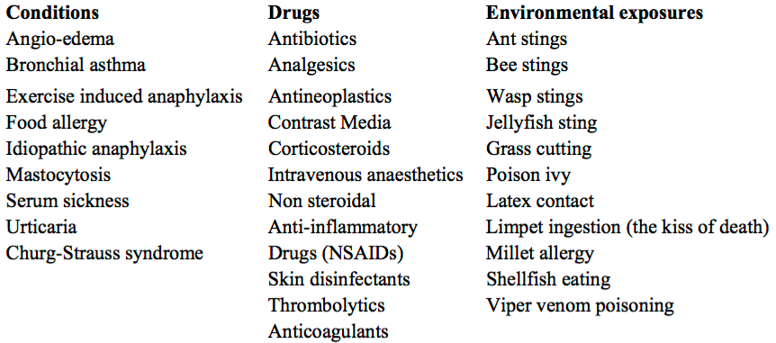
Causes of Kounis Syndrome


Treatment
•   Steroids
•   Anti-histaminics (H1 and H2 blockers)
•   Vasodilators (NTG) and routine ACS treatment

•   Adrenaline is the drug of choice and can save lives in anaphylaxis, but in Kounis syndrome there is a chance of aggravating ischemia and worsen coronary vasospasm. Despite this risk, in severe reactions, adrenaline should be given as wide spread manifestations of Anaphylaxis are life-threatening. Sulfite free adrenaline is recommended when available. Glucagon may be considered for those who take beta blockers and do not respond to adrenaline. Fentanyl shows a slight mast cell activation as compared to morphine and should be the drug of choice when opioid analgesia is necessary.


Posted by:


     Lakshay Chanana
     
     Speciality Doctor
     Northwick Park Hospital
     Department of Emergency Medicine
     England

     @EMDidactic


References:

  1. Kounis NG, Zavras GM. Histamine-induced coronary artery spasm: the concept of allergic angina. Br. J. Clin. Pract.45,121–128 (1991).
  2. Kounis NG, Zavras GM. Allergic angina and allergic myocardial infarction. Circulation94,1789 (1996).
  3. Kounis NG, Grapsas GM, Goudevenos JA. Unstable angina, allergic angina and allergic myocardial infarction. Circulation100,e156 (1999).

Sunday, March 12, 2017

Diffuse Esophageal Spasms (DES)

Diffuse Oesophageal Spasm, also called as "Prinzmetal Angina of the GI tract" is characterized by oesophageal contractions that are uncoordinated, simultaneous, or rapidly propagated. Usually, several segments of the esophagus contract simultaneously, preventing the propagation of the food bolus. 

Several patients are discharged from ED with a diagnosis of Non-Cardiac Chest pain or MSK pain when the ACS work up turns out to be negative. However,  Esphageal Spasm is not a ED based diagnosis. It often requires a trial of medications such Nitrates/CCBs or studies such as Manometry or Barium Swallow to reach a definitive diagnosis. Regardless, it is important for Emergency Physicians to be aware of this condition to arrange appropriate follow with Gastroenterologists. 


Symptoms:

  • Non-Exertional Retrosternal Chest Pain which frequently radiates to the back, and can be more severe than angina (May sound like dissection, pancreatitis, GERD, ACS)
  • Globus (ie, the sensation that an object is trapped in the throat)
  • Dysphagia, which is more consistent and reproducible during investigative studies
  • Regurgitation and Heartburn 
The spasms of the oesophageal muscles can lead to a feeling of food sticking, food obstructing, regurgitation, and chest pain. Symptoms may be worse with cold foods or drinks, but may improve with warm liquids.


Diagnosis:

It is reasonable to rule out ACS with ECGs/Troponins even when there is slightest of concern. Other helpful investigation are:
  • Bedside ECHO (Cardiac Contractility, RWMAs, Tamponade, Dilated Right heart, PTx)
  • CXR (Pneumonia/PTx, Dilated mediastinum)
  • Amylase (Pancreatitis)


The diagnostic modalities of choice for DES are barium swallow and esophageal manometry. Diffuse esophageal spasm has a characteristic appearance of multiple simultaneous contractions causing a corkscrew appearance with segmentation. It is important to remember that Barium Swallow will show this typical corkscrew pattern only if done during an episode of spasm. 


Treatment:
  • Calcium channel blockers and nitrates are first-line therapy. Other Treatment options: Sildenafil, Botulinum toxin, Diltiazem
  • Surgical Treatment - Myotomy/ Esophagectomy


Author:

              
     Lakshay Chanana
     
     Speciality Doctor
     Northwick Park Hospital
     Department of Emergency Medicine
     England

     @EMDidactic


Monday, November 7, 2016

Chasing Chest Pains - Risk Stratification and Non Specific Troponinemia

Chest pain is caused by a myriad of causes ranging from benign to life threatening, some of which can cause death within minutes or hours. While evaluating chest pain, ACS is always high up on our list of differential diagnosis and as Emergency Physicians, it is our responsibility to not only robustly identify ACS and but also avoid needless investigations and unnecessary admissions for those who can be safely discharged from the ED after risk stratification.


Here is an overview of ACS with breakdown of terminologies, key points about history/physical exam and biomarkers and specifics about what to do with a low risk ACS patient:

Classical ACS presents with:
  •             Heavy, aching or tight
  •             Central chest or left sided
  •             Not related to respiration or movement
  •             May radiate to arms, neck, or jaw


You will often see patients who have one or a few of these features but end up having a completely negative work up for ACS. Remember, the history is helpful only to risk stratify – not to confirm your diagnosis. Everyone perceives pain in a different way but history is the first step during evaluation and risk stratification. 

Atypical ACS
Atypical presentations of ACS are common, occurring in up to 1/3rd of patients, mostly in the elderly, diabetics and women. Advanced age, co-morbid factors, delay in diagnosis lead to the increased mortality in these populations.


Things you must ask/look for:

  • Radiation to both arms – Likely  ACS
  • Radiation to left arm – Likely ACS
  • Nausea / Vomiting – Likely ACS
  • SOB on exertion – Likely ACS
  • Associated with Sweating – Likely ACS
  • Hypotension – Likely ACS
  • S3 – Likely ACS
  • Describes as previous angina - Likely ACS


  • Pleuritic/ Positional/ Sharp Pain – Unlikely ACS (not impossible)
  • Tender on Palpation – Unlikely ACS (not impossible)


A good history helps in risk stratification. Don’t rule out ACS just based on the history alone. With the slightest of concern, get an ECG.


Risk Factors
Risk factors once again help us to risk stratify but just based on the absence of risk factors you cannot rule out ACS. Get worried if the history is concerning even if there are no risk factors at all. The next step is ECG.


And what if the history is concerning but ECG is normal?
An ECG showing ST depressions, TWI or STE is obviously concerning. Patients who present with chest pain with suspected cardiac ischemia based on the history but normal ECG should still undergo further diagnostic testing with cardiac markers before they can be confidently assigned to a low risk group.


What if the history is concerning with ongoing ischemic symptoms, but ECG is normal and troponin is not elevated?
This sounds very much like Unstable Angina. DO NOT SEND ANYONE HOME WITH CONCERNING AND PERSISTING SYMPTOMS DESPITE NEGATIVE TROPONINS.

Unstable Angina can occur when you are resting, sleeping, or during  little physical exertion. The pain may last longer than stable angina and rest or anti-ischemic medications usually do not help relieve it. USA can have an ischemic or normal ECG but should always have negative troponins by definition.

In contrast, Stable Angina is very predictable with Chest Pain on exertion that gets better on resting. Stable Angina us also relieved with anti-ischemic medications.


Beware of the Non-Specific Troponinemia AKA Troponinitis!

Troponins are the preferred and recommended markers of myocardial necrosis. Read more about troponins here. But the new generation hs troponins are extremely sensitive and thus less specific i.e you might end up getting a false positive elevated troponin leading to unnecessary admissions and work ups.  So if history is not suggestive of ACS but troponin is elevated – get a few more ECGs but do think of other possible causes of an elevated troponin such as:


With hs Troponins, we are now able to pick up even the minimal level of troponins in a patient’s system and thus those who were diagnosed as Unstable Anginas (Ischemic Chest Pain/ECG but normal troponin levels) a decade back are now labeled as NSTEMIs (Ischemic Chest Pain/ECG but a raised troponin). At this moment, although it is controversial but some experts do believe that Unstable Angina does not even exist anymore in this era of highly sensitive troponins.


ACS includes STEMI, NSTEMI, USA (not stable angina)
Patients with STEMI do not require troponin since their initial treatment is determined by their clinical presentation and ECG findings. Patients with STEMI are identified quickly, assigned a high risk category and have a well-defined treatment strategy (ie. urgent reperfusion with PCI or thrombolytics).


When does NSTEMI need immediate cathlab:
The ACC/AHA guidelines for NSTEMI recommend < 2 hour cath for:
  1. Refractory ischemia
  2. Ischemia with hemodynamic or electrical instability


If you are worried about a patient, get serial ECGs, send troponins and involve cardiology at the earliest.


Disposition of a Low Risk Patient – Slightly concerning history but non-ischemic ECG and negative enzymes. 

Here we are specifically talking about Unstable Anginas which can be further divided into two groups i.e negative troponin with ischemic ECG and negative troponin with a non-ischemic ECG. 

Current data shows that if patients have negative troponins with non-ischemic ECG, then prognosis is not bad even if they are diagnosed with unstable angina.  If they have unstable angina with an ischemic EKG, then they have a worse prognosis.

Note - if you see an Ischemic ECG – Get worried even when if the enzymes are normal

Low risk unstable angina with negative troponins can have:
Shared Medical Decision Making -  Do serial troponins and serial ECGs. Current evidence suggests a repeat troponin at hour 3 from initial EKG reduces potential miss rate from 1.7% to <1% at 30 days. Let them make this decision - ask them if they would want to get admitted or if they are happy to follow up as an out-patient with a week. 


Use Clinical Decision Making rules such as HEART/GRACE score to further risk stratify them and most important - Document medical decision-making and Clinical Decision rules  in the patient's record.


Read more on HEART SCORE on REBELEM.


Take Home:
  • A good history helps in risk stratification. Don’t rule out ACS just based on the history alone. With the slightest of concern, get an ECG.
  • It is okay to send troponins on your patients if you have some degree of concern but If there are no concerns eat all, then do not send troponins.
  • Patients who present with chest pain with suspected cardiac ischaemia based on the history but normal ECG should still undergo further diagnostic testing.
  • USA can have an ischemic or normal ECG but should always have negative troponins by definition.
  • Low Risk - Do Serial ECGs, Shared Decision Making, Clinical Decision Making Rules to further risk stratify and DOCUMENT the decision making in the medical record. 


Further Reading:
  • http://hqmeded-ecg.blogspot.co.uk/2014/04/unstable-angina-dr-braunwald-asks-if-it.html
  • http://hqmeded-ecg.blogspot.co.uk/2015/06/unstable-angina-still-exists-beware.html
  • https://blog.essentialsofem.com/2016/02/25/low-risk-chest-pain-adp-showdown-using-timi-vs-heart-pt-1-of-3-timi/


Author:

              
     Lakshay Chanana
     
     Speciality Doctor
     Northwick Park Hospital
     Department of Emergency Medicine
     England
     @EMDidactic