Monday, June 25, 2018

Singultus (Hiccups)

Pathophysiology of Hiccups
Hiccups or Singultus are involuntary, intermittent, spasmodic contractions of the diaphragm and intercostal muscles causing sudden inspiration that ends with abrupt closure of the glottis, making a classic sound. Hiccups are called 
Persistent if they last more than 48 hours and 
Intractable if they last more than 2 months. 


Hiccups during sleep suggest an organic cause

Exact pathophysiology of hiccups is not known. The reflex arc involves
1) the phrenic nerve, vagus nerve, sympathetic chain 
2) A central mediator
3) Phrenic nerve, glottis, and intercostal muscles. 

The central mediator is thought to involve the respiratory centers, phrenic nerve nuclei, reticular part of brainstem, and hypothalamus. Hiccups may result from stimulation, inflammation, or injury to one of the nerves of the reflex arc.

Causes of Hiccups (Consider the reflex arc for areas of possible etiologies)
  • Gastric distention
  • Carbonated drinks
  • Aerophagia
  • Sudden changes in GI or ambient temperature
  • EtOH, Smoking
  • Sudden excitement or stress
  • Vagus Nerve and irritation of its branches irritation - FB ear, Pharyngitis, Neck Masses, Esophagitis, Asthma, Thoracic Aorta Aneurysm, Stomach distention, AAA, Cholecystitis, Pancreatitis,  Appendicits
  • Goiters, mediastinal masses, tumors can tickle the Phrenic nerve
  • Diaphragm Irritation: hiatal hernia, GERD, subphrenic abscess, Myocardial ischemia, Pericarditis, manipulation during surgery
  • CNS disorders: Anything that disrupts the inhibitory hiccup reflex. Can be vascular (Brainstem CVA), infectious, or structural. Consider MS, hydrocephalus, syringomyelia, masses.
  • Toxic/metabolic: Can see with anesthesia, alcohol, HypoNa/Ca/K, Uremia
  • Psychogenic


ED Management
A thorough history and physical.  Consider investigations beased on suspected etiology and try simple remedies - Breathing into bag, swallowing sugar, forcible traction on the tongue, biting a lemon, breath holding, fright, noxious odors, ice water gargles, NG tube stimulation. Intractable hiccups may require phrenic nerve ablation. 

  • Medications: Address underlying cause. Options include: Chlorpromazine, Metoclopramide, Gabapentin, Baclofen, Haloperidol, low dose Ketamine. 

page1image2144
Take Home:
  • Hiccups are usually benign and involve a reflex arc involving the phrenic and vagus nerves, diaphragm, glottis, and intercostals.
  • Work-up should be reserved for patients with intractable or persistent hiccups.
  • Treatment modalities are varied, ranging from anecdotal techniques to phenothiazines, dopamine antagonists, anticonvulsants. 


References:
  1. Cymet. Retrospective analysis of hiccups in patients at a community hospital from 1995-2000. J Natl Med Assoc, June 2002
  2. Lierz and Felleiter. Anesthesia as therapy for persistent hiccups. Anesth Analg. Aug 2002.
  3. Thoma, et al. Behavioral treatment of chronic psychogenic polydipsia with hyponatremia: a unique case of polydipsia in a primary care patient with intractable hiccups. J Behav Ther Exp Psychiatry. 2001 Dec
  4. Dobelle. Use of breathing pacemakers to suppress intractable hiccups of up to thirteen years duration. ASAIO J. 1999 Nov-Dec;45(6):524-5.
  5. Pelag and Pelag. Case report: sexual intercourse as potential treatment for intractable hiccups. Can Fam Physician. 2000 Aug;46:1631-2.

Posted by:

              
     Lakshay Chanana
     
     Speciality Doctor
     Northwick Park Hospital
     Department of Emergency Medicine
     England

     @EMDidactic


Monday, June 18, 2018

Headache in children - ED management

Headache is a rare chief complaint in children but more frequently seen in adolescents. ED evaluation should answer the following the questions:
  • Primary or Secondary Headache?
  • Needs Investigations or Imaging?
  • Need for follow up with GP?
Primary headaches (Disabling): Migraine, Tension Headache, Cluster headache (and other trigeminal autonomic cephalalgias); and other primary headaches (such as cough or exertional headache). They are thought to be due to a primary malfunction of neurones. Primary headaches are typically self-limited.

Secondary headaches (a/w Morbidity and Mortality): May show up symptomats of some underlying pathology, e.g. from raised intracranial pressure and space-occupying lesions. Headaches due to a SOL are worse when lying down and morning vomiting is characteristic. There is often a change in mood, personality or educational performance. Causes of secondary headache include brain tumors, vascular malformations, and intracranial abscesses; craniofacial problems, such as sinusitis, dental abscesses, or otitis; systemic disorders, such as lupus cerebritis; and exposure to toxic substances, such as carbon monoxide, lead, or cocaine. Other features suggestive of a space-occupying lesion are i.e Red Flags for Headache:

  • Visual field defects – from lesions pressing on the optic pathways, e.g. craniopharyngioma (a pituitary tumour)
  • Cranial nerve abnormalities causing diplopia, new-onset squint or facial nerve palsy. Abducens nerve has a long intracranial course and is often affected when there is raised pressure, resulting in a squint with diplopia and inability to abduct the eye beyond the midline. It is a false localising sign. Other nerves are affected depending on the site of lesion, e.g. pontine lesions may affect the VIIth (facial) cranial nerve and cause a facial nerve palsy.
  • Abnormal gait (Cerebellar Lesions)
  • Torticollis 
  • Bradycardia
  • Confusion, Seizures
  • Growth failure, e.g. craniopharyngioma or hypothalamic lesion
  • Papilloedema 
  • Cranial bruits – may be heard in arteriovenous malformations 
  • Recent change in personality, behaviour or educational performance
  • Headache that wakes up a child (different from headache on awakening, not uncommon in migraine)
Headache coinciding with the onset of fever suggests inflammation - sinusitis, pharyngitis, otitis, or meningitis), or may be associated with a more general viral syndrome. The severity of a headache is not helpful to determine the cause. Patients with tension headaches can com- plain of terrible pain, whereas a child with a brain tumor may complain of mild to moderate pain. Nonetheless, complaints of very intense pain should be taken seriously and assessed in context with other historical elements.


The most common causes of headache in chidren are viral and respiratory illnesses and migraneous headaches. A migraine that lasts >72 hours is known as status migrainosus


  • The mainstay of management is a thorough history and examination. Imaging is unnecessary in the absence of any Red Flag features.
  • Among children with intracranial diseases, most are unable to indicate the location of the pain or had an occipital headache. 
  • Positional preferences --> space- occupying lesions
  • Abdominal pain, nausea, or vomiting--> May be seen in migraine 
  • Cluster headaches can be associated with multiple ipsilateral symptoms
  • Headache with effortless vomiting but no GI complaints --> suggests elevated ICP
  • Family history is commonly seen in migraneous headaches

Examination findings may be normal even if the history suggests a secondary headache. A careful neurologic examination with attention to cranial nerves, gait, strength, and mental status is essential to exclude secondary headache. There is a very low incidence of positive findings on neuroimaging studies in children with headache who have normal findings on physical examination.

Imaging CT or MRI - NCCT is sufficent for most cases that require imaging. CT with contrast is needed when you suspect brain abscess but normal head CT does not exclude an intracranial mass because the posterior fossa is not well visualized. The posterior fossa is the most common location of brain tumors in children, and MRI of the brain provides better visualization of the posterior fossa. MRA/MRV are useful when vascular malformations or dural sinus thrombosis is suspected. 


Treatment
Do not use narcotics for primary headaches. Long-term use of narcotics can change the pain-modulatory system leading to more intense pain. 

Options: 
  • Primary Headaches - Fluid bolus/NSAIDs, Stemetil or Metoclopermide. Metocloperamide and Stemetil can cause extrapyramidal reactions, which are relieved by diphenhydramin, Triptans High Flow Oxygen can also be used to treate Cluster HA.  
  • Secondary Headaches need symtom control with Analgesics and further evaluation. 


Take Home:
The mainstay of management is a thorough history and examination. Imaging is unnecessary in the absence of any Red Flag features.


Posted by:

              
     Lakshay Chanana
     
     Speciality Doctor
     Northwick Park Hospital
     Department of Emergency Medicine
     England

     @EMDidactic


Monday, June 11, 2018

Children with tLOC

Children presenting with syncope usually suffer from benign causes and about 80% constitute vasovagal or neuro cardiogenic syncope. From an etiological perspective, syncope in children can be divided into four likely causes:

  1. Cardiovascular (tachydysrhythmias, bradydysrhythmias, outflow obstruction, and myocardial dysfunction)
  2. Neurologic 
  3. Metabolic 
  4. Other Benign entities (Vasovagal - reduced venous return)

A prodrome of warmth, nausea, lightheadedness, and a visual gray-out is indicative of neurocardiogenic syncope. Routine laboratory studies are not needed in such cases.

Involuntary motor movements due to transient cerebral hypoxia may occur in syncopal events which may be reported as fits by laypersons. Syncope related myoclonus is generally seen after loss of consciousness (seizure-related movements are seen prior to the loss of consciousness), less rhythmic nature, and are of shorter duration. 


Red Flags for Pediatric Syncope:
  • Onset with Exertion
  • History of Cardiac Disease
  • Heart Murmur on examination
  • Family h/o Sudden Deaths or arrhythmias
  • Associated chest pain/palpitations
  • Syncope in Recumbent position
  • Recurrent episodes
  • Use of Cardiac medications
  • No prodrome  
  • Prolonged LOC

Exertion before a syncopal event increases the suspicion of structural heart disease, specifically cardiac outflow obstruction. 


Investigations
  • ECG - Remember, ECG is a snapshot and some patients with an underlying arrhythmia may have normal ECG
  • Selection of other laboratory tests should be guided by clinical suspicion. Blood tests are required as indicated by history. Consider performing a serum alcohol level, a urine drug screen and a beta hCG in adolescents. 
  • ECHO - Done for those with known cardiac disease, abnormal heart sounds, abnormal cardiac murmurs, evidence of cardiac chamber enlargement, or repolarization abnormalities on ECG, or other features that suggest myocardial dysfunction.
  • EEG -  Low diagnostic yield and not needed routinely.


A detailed history, physical exam, and ECG have a 96% sensitivity for detecting cardiac syncope.

Things to look for in ECG:

  • Brady and Tachyarrythmias (AV Blocks, SVT, AF, VT)
  • Brugada (IRBBB, STE V1-V3)
  • QTc (Long and Short)
  • WPW (triad - Wide QRS, Short PR, Delta wave)
  • HOCM (High LV Voltage, dagger like q waves, TWI)
  • ARVD (epsilon wave)
  • Chamber Enlargement 
  • Septal Defects (Crochetage Pattern for OS ASD)


Treatment
Treatment is targeted to specifically identified causes of the syncopal event; 80% of the time, this will be neurocardiogenic syncope, and treatment for these patients includes reassurance and oral fluids. Children with a normal ECG, full recovery and no cardiac risk factors or exercise-induced symptoms may be safely evaluated as outpatients.


Take Home:
  • A detailed history, physical exam, and ECG have a 96% sensitivity for detecting cardiac syncope.
  • There are no routine blood tests - request bloods based on history and physical.
  • Know what to look for in the ECG in patients presneting with syncope.


Posted by:

              
     Lakshay Chanana
     
     Speciality Doctor
     Northwick Park Hospital
     Department of Emergency Medicine
     England
     @EMDidactic


Monday, June 4, 2018

Acute Urinary Retention

Acute urinary retention (AUR) is commonly encountered condition in the emergency department. It is the inability to void voluntarily despite a distended bladder that often leads to agitation or altered mental status in the elderly who are on several medications. Classical patient with AUR is an elderly man with BPH. 


Hypertension or tachycardia may be transient and may resolve after bladder decompression. 


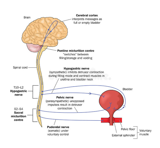
Higher Centres and Receptors involved in Micturation
Micturition involves coordination of high cortical neurologic (sympathetic, parasympathetic, and somatic) and muscular (detrusor and sphincter smooth muscle) functions.


Urinary Retention requires both relaxation of the detrusor muscle (through Î²-adrenergic stimulation and parasympathetic inhibition) and contraction of the bladder neck and internal sphincter (through Î±-adrenergic stimulation). In contrast, Urination requires contraction of bladder detrusor muscle (by cholinergic muscarinic receptors) and relaxation of both the internal sphincter of the bladder neck and the urethral sphincter (throughα-adrenergic inhibition). 

Common Causes of Urinary Retention

  • BPH
  • Prostate Cancer, Phimosis, Paraphimosis
  • Meatal Stenosis
  • Prostatitis
  • Medications (Anticholinergics, Antihistaminics, Antipsychotics, BZDs etc.)

Females presenting with AUR should undergo neurologic examination and a pelvic examination to detect possible inflammatory lesions or pelvic masses. 

AUR is a clinical diagnosis but bedside US can be used as an adjunct. Prolonged obstruction may result in impaired renal function and electrolyte imbalance. Thus, renal function studies and Potassium should be checked for those with prolonged retention. Formal abdominal imaging and urodynamic studies can be deferred as an out-patient if the patient appears clinically well. 

A thorough history is required to find the cause/precipitant of Urinary Retention

ED management is limited to bladder decompression with urethral catheterization or suprapubic catheterization (if urethral cath fails). Alpha-blockers can be prescribed during discharge to relax the uretheral muscles. 


Disposition and Indications for admission

Admit in case of:
  • Significant Post Renal Failure
  • Post Obstructive Diuresis
  • Frank Hematuria
  • Clot Retention 
  • Sepsis
Majority of patients with AUR are discharged home after bladder decompression and Urology Clinic review. It is paramount to educate them about catheter care to avoid accidental displacement/removal of the catheter leading to urethral injury. Reg flags include - fever, abdominal pain, catheter blockage, or penile pain. Tho who complain of a sense of urgency despite being on foleys can be treated with oxybutynin, 2.5 milligrams twice/thrice daily. 

Posted by:

              
     Lakshay Chanana
     
     Speciality Doctor
     Northwick Park Hospital
     Department of Emergency Medicine
     England

     @EMDidactic