Monday, December 25, 2017

Post-Partum Haemorrhage

Postpartum hemorrhage that occurs within the first 24 hours of delivery is called as primary postpartum hemorrhage. The main causes of primary postpartum haemorrhage are:
  • Uterine atony (TONE)
  • Retained placental fragments (TISSUE)
  • Lower genital tract lacerations (TRAUMA)
  • Uterine rupture (Click here to read more)
  • Uterine inversion (requires repair under general anesthesia)
  • Hereditary coagulopathy (THROMBIN)

Secondary postpartum hemorrhage occurs after the first 24 hours and up to 6 weeks postpartum. Common causes of secondary postpartum haemorrhage are:
  • Failure of the uterine lining to sub-involute at the former placental site
  • Retained placental tissue
  • Genital tract wounds
  • Uterogenital infection
Causes can be remembered as TONE, TISSUE, TRAUMA, THROMBIN


Risk Factors fro PPH
  • Primipara or Grandmultipara 
  • Previous PPH
  • Pre-eclampsia
  • Prior CS
  • Placenta Previa
  • Cervical or Uterine trauma
  • Fetal Wt >4.5Kgs
  • Prolonged 3rd stage
Excessive blood loss in the postpartum period is defined as a 10% drop in the hematocrit, a need for transfusion of packed red blood cells, or volume loss that causes symptoms of hypovolemia. The hematologic changes of pregnancy can mask the typical symptoms of hemorrhage, and the first sign may be only a mild increase in pulse rate.


Resuscitation
  • ABC
  • IV Access x 2
  • Fluid Resuscitation 
  • Involve OBGYN ASAP
  • Keep them warm (Prevent the deadly triad of hypothermia, coagulopathy and acidosis)
  • Bimanual uterine massage - place a fist in the anterior fornix and compress the uterine fundus against the hand in a suprapubic location 
  • Uterotonics 
Oxytocin: 10U IM or 20-40 units in NS over 1 hour 
Carboprost: 250mcg IM q30min (up to 2mg if needed), Avoid in HTN, Asthma
Misoprostol: 1000mcg PR
Methylergonovine: 0.2mg IM (up to 5 doses q2-4h), Contraindicated in HTN/Pre-eclampsia
  • Consider Tranexamic Acid for critically ill 
  • Look for evidence of trauma, uterine inversion and uterine rupture
  • Inspect for missing placenta fragments 
  • Arrange blood products (Packed Cells, FFP and Cryo if in DIC)
  • Intrauterine balloon tamponade using Bakri balloon or Rusch catheter if uterine atony is the only or main cause of haemorrhage
  • Move to OR for hysterectomy or Uterine Artery Ligation


Other advanced care methods:
  • Interventional Radiology for Uterine Artery Embolisation
  • REBOA as a temporary measure 
Take Home:

  • Keep them warm (prevent Hypothermia, Coagulopathy and Acidosis)
  • Remember the 4 causes - TONE, TISSUE, TRAUMA, THROMBIN
  • Involve OBGYN ASAP


References and Further Reading:
  1. Tintinai EM 8th edition
  2. Shakur H, Elbourne D, Gülmezoglu M, Alfirevic Z, Ronsmans C, Allen E, Roberts I. The WOMAN Trial (World Maternal Antifibrinolytic Trial): tranexamic acid for the treatment of postpartum haemorrhage: an international randomised, double blind placebo controlled trial. Trials. 2010 Apr 16;11(1):40.
  3. https://www.rcog.org.uk/en/guidelines-research-services/guidelines/gtg52/

Posted by:

              
     Lakshay Chanana
     
     Speciality Doctor
     Northwick Park Hospital
     Department of Emergency Medicine
     England

     @EMDidactic


Monday, December 18, 2017

Renal Colic

Acute Pain is one of the key reasons why patients present to Emergency Department and we should be experts at managing any sort of acute pain. Renal Colic is one such pathology that presents with sudden onset intermittent severe crampy flank pain a/w nausea and vomiting. NSAIDs are the drugs of choice as they inhibit prostaglandin synthesis and result in relaxation of ureteral spasm and decrease of renal capsular distension and associated pain. Here is a quick review of Renal Colic:


Risk Factors for Renal Stones
  • Obesity
  • Diabetes
  • Metabolic Abnormalities
  • Hyperparathyroidism
  • Immobilisation
  • Excess intake of meat and Na
  • Gout
  • Inflammatory Bowel Disease
  • Family History


Up to 15-30% patients with Nephrolithiasis may not show blood in urine. Do not exclude tis diagnosis based on the absence of hematuria. 


Things that you should not miss (Mimics):
  • Dissection/Aneurysm (most common misdiagnosis given to patients with a rupturing or expanding abdominal aortic aneurysm)
  • Renal Infarct
  • Pyelonephritis
  • Biliary Colic
  • Pancreatitis
  • Diverticulitis
  • Ovarian Torsion
  • Ectopic Pregnancy
  • Lower Lobe Pneumonia
  • Testicular Torsion
  • Herpes Zoster

Investigations:
  • Urine Dip (look for infection)
  • Full Blood Count (WCC is elevated due to stress demargination)
  • Uric Acid, CA (Helps in further evaluation as an out-patient)
  • Renal Function (Normal creatinine does not rule out obstruction)
  • Imaging (CT KUB, Bedside USG, Formal USG) - read more on imaging at aliem
  • Beta hCG
Consider adding amylase, LFT, CXR if history or examination findings are atypical. 


Management
  • Pain Relief (IM/IV/PR NSAIDs, Opioids, Antispasmodics are of uncertain benefit)
  • Anti-emetics (Metocloperamide)
  • Medical Expulsion Therapy (No proven benefit. Prescribe only for >5mm distal ureteric stones)
  • Antibiotics if febrile, systemically unwell (WCC is elevated due to stress demargination)
IV Fluids do not expedite stone expulsion


Consult Urology in cases of:
  • Refractory Pain
  • Obstruction and Acute Renal Failure
  • Urosepsis
  • Advanced age and co-morbidities
  • Solitary/Transplanted Kidney
  • Pregnancy

Take Home
  • Look for risk factors and potential mimics (aortic dissection and renal infarct)
  • Do not rule out kidney stones based on the absence of hematuria
  • Provide pain relief and arrange follow up 

Further Reading
  1. Core EM - Renal Colic
  2. REBEL EM - Does Use of Tamsulosin in Renal Colic Facilitate Stone Passage?
  3. Golzari, S. E., Soleimanpour, H., Rahmani, F., Zamani Mehr, N., Safari, S., Heshmat, Y., & Ebrahimi Bakhtavar, H. (2014). Therapeutic Approaches for Renal Colic in the Emergency Department: A Review Article. Anesthesiology and Pain Medicine4(1), e16222. http://doi.org/10.5812/aapm.16222
  4. Stewart A, Joyce A. Modern management of renal colic. Trends in Urology & Men's Health. 2008 May 1;13(3):14-7.

Posted by:

              
     Lakshay Chanana
     
     Speciality Doctor
     Northwick Park Hospital
     Department of Emergency Medicine
     England

     @EMDidactic


Monday, December 11, 2017

ED Dementia Screening

Dementia

Diagnosing dementia constitutes "memory loss" in addition to one of the following:
  • Apraxia (difficulty executing motor tasks)
  • Aphasia (language impairment)
  • Agnosia (difficulty recognising familiar objects)
  • Loss of executive function (planning, organising)
More than 50% of the patients presenting to Emergency Department have dementia and in about 3/4 the of the patients, the diagnosis is not yet established. Patients with a new diagnosis of dementia may have several implications on continuation of care as dementia can be associated with poor drug compliance, self-neglect, depression, suicidal risk. It is paramount to discuss these concerns with social services prior to discharging these patients. 


Mini-Cog Assessment 

  1. Ask the patient to repeat and then remember 3 unrelated words (ex. apple table tree)
  2. Give the patient a piece of paper with a circle on it
  3. Instruct the patient to place numbers on it to represent the face of a clock. After the patient puts the numbers on the clock face, ask him to draw the hands of the clock to read any specific time
  4. Ask the patient to repeat the 3 previously presented words
Scoring Mini-Cog Assessment (Minimum score 0, Maximum 3)
Give 1 point for each recalled word 

  • 0 - positive screen for dementia
  • 1 or 2 with an abnormal clock -  positive screen for dementia
  • 1 or 2 with a normal clock - negative screen for dementia
  • 3 - negative screen for dementia
    Patients who are called "poor historians" often have underlying dementia. If you suspect dementia in ED, then arrange follow up care with Neurology for thorough assessment. 


    Discharge checklist for Dementia-

    • Ask them their home address and how will they get there?
    • Sucidal Thoughts/Depression screen? 
    • Carers/Next of kin informed?


    Further Reading:

    Borson, S., Scanlan, J. M., Chen, P. and Ganguli, M. (2003), The Mini-Cog as a Screen for Dementia: Validation in a Population-Based Sample. Journal of the American Geriatrics Society, 51: 1451–1454. doi:10.1046/j.1532-5415.2003.51465.

    Posted by:

                  
         Lakshay Chanana
         
         Speciality Doctor
         Northwick Park Hospital
         Department of Emergency Medicine
         England

         @EMDidactic


    Monday, December 4, 2017

    Acute Abdomen in Elderly


    Working up an elderly population presenting with abdominal pain is always a challenging task. Almost always there are chances of potential mishaps due to delayed or missed diagnosis. Data suggests that abdominal pain is the most common ED presentation and the fourth most common complaint among elderly and nearly half of elderly patients with abdominal pain will require admission, and 1/3 will require  a surgical intervention. This makes it an important topic for us to be familiar with. Usual issues related to geriatric population (pharmacologic, social, cognitive) make evaluation extremely difficult. Click here to read more about how is elderly population different.
     

    Key Issues:


    • Present later in their disease course
    • Present with vague symptoms (Only 1/6 present with textbook appendicitis presentation)
    • Difficult Communication (hearing, visual, cognitive)
    • Underreport symptoms (Poor pain perception, fear of hospitals and medical interventions)
    • Physical exam has limited utility - Atrophy of abdominal wall musculature diminishes rebound and guarding
    • Medications blunt or alter their response to disease
    • Less likely to develop fever or leukocytosis
    In A&E, we should have a low threshold to image elderly due to high likelihood of surgical illness and unreliable physical examination. Ideally, CT is the imaging of choice. Plain films are of limited utility. Due to unreliable history, misleading examination findings and unpredictable nature and course of the illness, admission for observation should be considered if no confusion is reached after ED work up. . 

    Common Geriatric Abdomen Pathologies

    Mesenteric Ischemia
    • Risk factors include Atrial Fibrillation, Heart Failure. 
    • Pain out of proportion to examination. Perform a quick assessment and come up with a plan. Involve your Surgical colleagues and Radiologists ASAP.
    • May present with normal vital signs and laboratory values. Do not overly on White cell count and lactate. Lactate and EBC both are non-specific. 
    • Imaging of choice is CT with IV contrast.
    •  Rx them with Pain Relief, Fluids and electrolytes, NG Tube for gut decompression, Foleys Catheterisation , Broad Spectrum Antibiotics and immediate surgical consultation. 


    Aortic Diseases (AAA, Dissection)

    • Be cautious whenever you attribute flank/abdo pain to Renal Stones in elderly.
    • AAA May present with weakness, dizziness, uneasiness or syncope
    • Classic presentation is pain abdomen, pulsalitle mass and hypotension
    • Use bedside USG to measure aorta and also look for a dissecting flap
    • Maintain BP enough to perfuse brain and do not give too much fluids
    • Arrange blood for transfusion and involve vascular surgeons ASAP


    Gall Bladder Disease and Pacreatitis


    • Perforation, gangrene, emphysematous cholecystitis, ascending cholangitis, gallstone ileus, choledocholiathisis, and gallstone-induced pancreatitis are all more prevalent 
    • More than 50% with acute cholecystitis will lack nausea, vomiting, or fever. Leukocytosis may be absent in 30% to 40% of those with acute cholecystitis.31 Evaluation by ultrasound may be less helpful given the increased prevalence of acalculous cholecystitis as well as cholodocolithiasis and delay in surgery may result in an increased mortality.
    • More than 50% cases pancreatitis and elderly are due to Gall Stones. 


    Small Bowel Obstruction


    • Classical symptoms are not seen early in the course of disease
    • May present with diarrhoea due to hyperperistalsis distal to the obstruction point
    • Gallstone disease may contribute to 25% of bowel obstructions in elderly


    Diverticulitis


    • Diverticular bleeding is one of the most common causes of lower GI bleeds
    • Acute diverticulitis occurs when the diverticula become obstructed by fecal matter, resulting in lymphatic obstruction, inflammation, and perforation. 
    • Usually presents with LLQ pain, with or without bloody stools, nausea, and fever but 1/3rd of the geriatric presentations of acute diverticulitis do not have abdominal tenderness on examination. Once the acute phase resolves, endoscopy should be performed to rule out carcinoma after an acute episode of diverticulitis. 
    • Most can be managed medically, with antibiotics, intravenous fluids, and bowel rest. If there are larger perforations or abscess formation, surgery or percutaneous drainage may be indicated.

    Large Bowel Obstruction

    • Common causes are diverticulosis and malignancy. 
    • Classically present with abdominal pain, vomiting, and constipation/obstipation, 
    • Often diagnosed late in their course 
    • Sigmoid and cecal volvulus account for a smaller subset of large-bowel obstructions, but more often requires emergent surgical intervention. 
    • Sigmoid volvulus, causing close to 80% of volvuli, causes a more gradual onset of pain, whereas cecal volvulus presents more acutely.
    • Sigmoid volvulus can often be decompressed with a rectal tube, sigmoidoscope, or barium enema, whereas cecal volvulus requires surgical repair. Volvulus of either site is at risk for perforation and should be decompressed urgently. 
    • Functional impairment and decreased motility of the GI tract can lead to acute colonic pseudo-obstruction, or Ogilvie syndrome i.e. functional obstruction of the GI tract. This is more commonly seen in elderly and debilitated patients. Treatment is conservative medical management. Neostigmine can be very effective but requires careful monitoring due to potential of bradycardia. 


    Constipation


    • May be associated with fecal impaction and fecal incontinence. Fecal impaction can cause mucosal ulceration, bleeding, and anemia. 
    • Often caused by Medications, comorbidities, inactivity, and decreased gastric-emptying time and GI malignancies 
    • Perform a rectal examination should be performed on all patients with constipation to rule out mechanical obstruction of stool.
    • Treat chronic constipation with dietary and activity changes, followed by bulk laxatives and warm water enema


    Malignancy


    • Ask for h/o unintentional weight loss, night sweats, and fatigue, hematuria, hematochezia. 
    • May also present with peritonitis, ascites, perforation, obstruction, or abdominal mass

    Appendicitis
    • Difficult to diagnose in elderly patient and missed in more than 50% cases. The classic presentation is rarely seen. Literature now supports the use of non-contrast CT imaging for suspected appendicitis.
    • Elderly have a higher risk for mortality and morbidity following appendectomy. 


    Extra-Abdominal Causes


    • Congestive heart failure
    • Acute Coronary Syndromes
    • Pneumonia, Pulmonary embolism
    • Prostatitis 
    • Urinary retention/infection (antihistamines, anticholinergics, technical obstruction, Pyelonephritis)
    • Herpes zoster involving thoracic dermatomes 
    • Rectus sheath hepatomas if they are on anticoagulants

    Take Home

    • Think of Vascular causes of pain abdomen in elderly (AAA, Dissection, Bowel Ischemia)
    • GB disease (Cholecystitis, Cholangitis, Pancreatitis) and Bowel Obstruction are common in geriatric population
    • Do a rectal exam to assess prostate, look for blood/malena
    • Do a broad work up including lab tests and have a low threshold for imaging and admission for observation


    Further Raeding: 
    Leuthauser A, McVane B. Abdominal pain in the geriatric patient. Emergency Medicine Clinics. 2016 May 1;34(2):363-75.


    Posted by:

                  
         Lakshay Chanana
         
         Speciality Doctor
         Northwick Park Hospital
         Department of Emergency Medicine
         England

         @EMDidactic


    Monday, November 27, 2017

    Patients requiring "medical clearance"

    Majority of the disease burden that we see in Emergency Departments includes Geriatric pathologies and Psychiatric issues. Whenever Psychiatry evaluates any patient, they always ask for "medical clearance" and want us to mention this specifically on the chart. I often come across patients who visit Emergency Department more than I do as an A&E physician. Regardless, they go through this process of "medical clearance" every time. 

    Frequently, it gets difficult to document "medically cleared". For instance, when psych patients have slightly high blood glucose but do not need anything besides some IV fluids or they might come up with a BP of 180/90 due to agitation. Additionally, getting a precise drug history can be a challenge in this subgroup of patients as they frequently take several medications for underlying chronic illnesses. It can be problematic if we document medically cleared on the chart and I believe "No acute medical concerns at this point or Fit for Psychiatric assessment" is a better way to clear these patients. Medical evaluation is used to determine whether the patient has a medical condition that is causing or exacerbating the psychiatric illness. 

    At any cost we do not want to miss an underlying medical pathology. Things that we need to look for to r/o organic cause of Psych symptoms:
    • ABCs as always
    • Vital Signs (PR, BP, RR, Temp, SpO2, Glucose) - Do not ignore abnormal VS
    • Pupils and Skin exam (pick cues for toxidromes)
    • Focused History (Next of Kin/Paramedics) and Physical exam
    • Focus on underlying Psych issues (Drugs/Alcohol, Sexual, Suicidal, Homicidal, Social aspects)
    • Medications and Co-Morbidities 
    • Document Mental Exam - Appearance, Behaviour, Cognition, Speech, Mood, Insight, Thoughts, Hallucinations

    Most of them do not require blood tests. Bloods are done in those with:
    1. New-onset psychiatric complaints
    2. Abnormal vital signs
    3. Elderly
    4. Known co-morbid conditions. 

    Even Urine Drug Sceen is not  required routinely for all. Most of them will tell you what drugs they have recently taken. Also remember, UDS may have false positive and negatives which can be misleading.


    Signs suggesting underlying medical pathology are:
    • Abnormal Vital Signs
    • Clouded Consciousness 
    • Age >40 with no previous Psych issues
    • Focal deficits on exam
    • Abnormal Physical examination 
    • Visual Hallucinations 

    Visual hallucinations can also occur in psychiatric illnesses but assume medical pathology until proven otherwise


    Take Home:

    • Spend more time on history and exam rather than doing bloods on everyone
    • Do not ignore abnormal Vital Signs
    • Look at issues that often co-exist with Psych illnesses - Drugs/Alcohol, Sexual, Suicidal, Homicidal, Social aspects

    Further Reading:

    • Korn CS, Currier GW, Henderson SO: Medical clearance of psychiatric patients without medical complaints in the emergency department. J Emerg Med 18: 173, 2000. 
    • American Psychiatric Association: Practice guideline for psychiatric evaluation of adults. Am J Psychiatry 152: 63, 1995. 
    • Korn CS, Currier GW, Henderson SO: “Medical clearance” of psychiatric patients with- out medical complaints in the emergency department. J Emerg Med 18: 173, 2000. 
    • Olshaker JS, Browne B, Jerrard DA, Prendergast H, Stair TO: Medical clearance and screening of psychiatric patients in the emergency department. Acad Emerg Med 4: 124, 1997. 
    • Broderick KB, Lerner B, Mccourt JD, Fraser E, Salerno K: Emergency physician practices and requirements regarding the medical screening examination of psychiatric patients. Acad Emerg Med 9: 88, 2002. 


    Posted by:

                  
         Lakshay Chanana
         
         Speciality Doctor
         Northwick Park Hospital
         Department of Emergency Medicine
         England

         @EMDidactic


    Monday, November 20, 2017

    Getting better at diagnosing Delirium


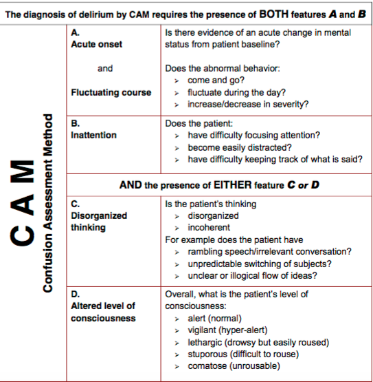
    Delirium can be defined as an acute confusional state caused due to medical or pharmacological triggers. Up to 10% of all older patients in EDs have delirium and only about one-third of them are recognizedAs frontline healthcare providers, it is essential to recognize delirium as the symptom of a life-threatening underlying medical or surgical condition. The consequences of a missed delirium can be disastrous as they can bounce back with florid sepsis, trauma, seizures. In elderly, delirium may be the only sign of an underlying infection (Pneumonia, Sepsis, ACS, Abdominal infection, Intra-cerebral event). Delirium is often multifactorial in etiology and each cause should be investigated. 

    DSM IV defines delirium as:
    • Disturbance of consciousness with reduced ability to focus, sustain, or shift attention
    • Change in cognition that is not better accounted for by a pre-existing, established, or evolving dementia
    • Development over a short period of time (usually hours to days) and disturbance tends to fluctuate during the course of the day
    • There is evidence from the history, physical exam, or lab findings that the disturbance is caused by the consequences of a general medical condition

    This can be remembered as:

    A - Acute and fluctuating (Often reported by a family member or carers)
    I - Inattention (Inattention is a hallmark feature of delirium - Can test with months of the years backwards, WORLD or serial 7s)
    D - Disorganised thinking (incoherent, rambling, circumstantial, or vague. (irrelevant conversation, unclear or illogical flow of ideas, or unpredictable switching from subject to subject)
    A - Altered Mental Status (hyperactive, drowsy, stuporous, comatose)
      
    Causes of Delirium:
    • Systemic Diseases (Infections, ACS, Hepatic Enceph, Metabolic - Na/Glucose/Ca disturbances)
    • Medications (Anticholinergic, Antiparkinsonian, Antiemetics)
    • Withdrawal (Discontinuation or overdose of any medication, alcohol, sedative hypnotics)
    • CNS Pathology (CVA, Subdural hematoma, Meningitis, Seizures – non convulsive, Hypertensive encephalopathy)
    Infections are the most commonly encountered cause of delirium in elderly and medications are the most common reversible causes of geriatric delirium.

    Why is Delirium missed in ED? 
    We frequently assume that most older people have baseline cognitive impairment. Collateral History and paying attention to what caregivers say is crucial. Delirium can present in hypoactive, hypoactive or mixed form. Therefore, it does not always present as restlessness or agitation, in fact hypoactive delirium is the commonest. Using formal assessment methods is the best way to avoid such mishaps


    Managing Delirium
    Most delirious patients need admission unless we find a single, reversible cause and patient has good social support and someone to observe him at home. 

    Non-Pharmacologic 

    • Manage pain and avoid unnecessary restraints including catheters and non-essential monitor leads
    • Promote mobility and encourage to eat and drink
    • Address bowel and bladder function
    • Increase sensory stimulation (hearing aid, eye glasses)
    • Enhance orientation and familiar faces (family)
    • Minimize medication changes 
    Pharmacologic 
    Medications should be used if evaluation is prevented or if the patient gets severely agitated. Haloperidol remains the drug of choice (Not Benzodiazepines). Use BZD only in case of drug withdrawals. Use Haloperidol 0.5mg-1.0mg PO/IM/SC/IV every 30min to 1hr and frequently reassess. Haloperidol is a potent anti-psychotic with limited anti-cholinergic effects though it may cause problems in Parkinsonian patients because of increased extra pyramidal symptom.

    Preventing Delirium
    ED Stay > 8 hrs itself is a risk factor for delirium. High Risk groups include dehydrated patients, demented and restrained patients, those who were left in the corridor overnight, visually and hearing impaired. 


    Further Reading

    • Altered Mental Status in Older Emergency Department Patients - Wilber, Scott T. Emergency Medicine Clinics , Volume 24 , Issue 2 , 299 - 316 
    • The Acutely Confused Elderly Patient

    Posted by:

                  
         Lakshay Chanana
         
         Speciality Doctor
         Northwick Park Hospital
         Department of Emergency Medicine
         England

         @EMDidactic