Wellens' Syndrome
Wellens’ syndrome
was first described by Prof Hein J.J. Wellens in 1982. It can be best described as a
warning for an impending heart attack. As emergency physicians it is crucial for us to be able to recognise this ECG pattern. Recognition of this ECG pattern can potentially prevent the myocardial infarction.
It is important to understand that Wellens’ syndrome is not just an ECG sign alone but ECG signs in context with the clinical picture illustrated below.
Identification
with examples
There are 2 types
of Wellens’ waves:
- Wellens' with biphasic T waves and - Figure 1
- Wellens' symmetrically inverted and deep T waves – Figure 2

Figure 1 (image courtesy - Lifeinthefastlane)

Figure 2 (image courtesy: Lifeinthefastlane)
1. Deeply inverted or biphasic T waves in V2-V3 sometimes extending from V1 to V6
2. Isoelectric or slightly elevated ST segment (< 1mm)
3. No pathological precordial Q waves
4. Preserved precordial R wave progression
5. Recent history of Angina
6. ECG pattern in pain free state
7. Normal or slightly elevated serum cardiac markers
Clinical Importance
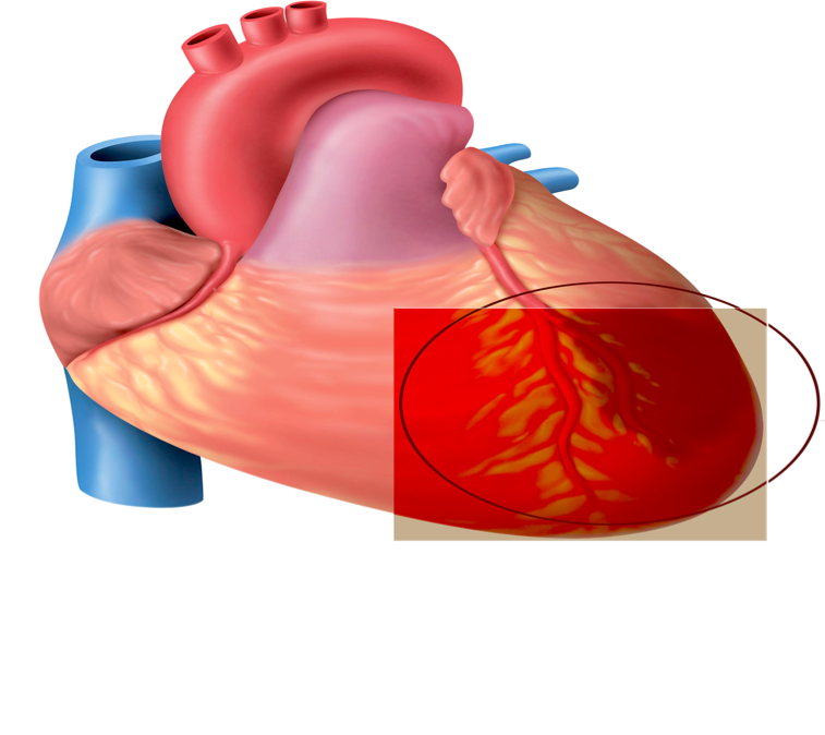
- Signifies critical left anterior descending artery occlusion and high risk for extensive myocardial infarction
- About 75% of patient with this ECG finding have AW STEMI within < 2 weeks.
- These patients may or may not have active chest pain at presentation and the cardiac enzymes may be normal or elevated
- These patients are not fit for stress tests as they are too high risk for a large myocardial infarct
Get cardiology involved and push for a timely angiography. Medical Management is usually ineffective for these lesions, these are best managed with PCI.
Key Points:
- Watch out for Wellen's waves routinely (esp biphasic T waves)
- This subset of patients may not have chest pain and normal cardiac biomarkers.
- Don't send them for a stress test.
- Discuss with Cardiology for urgent/emergent PCI
References
1. de Zwaan C,
Bar FW, Wellens HJ. Characterstic electrographic pattern indicating a critical
stenosis high in left anterior descending coronary artery in patients admitted
because of impending myocardial infarction. Am Heart J. 1982 Apr; 103: 730-6
2. Rhinehardt et al. Electrographic
manifestations of Wellens Syndrome.
Am J of Emergency Medicine 2002 Nov; 20(7):638-43
3. Liu Mao et al.
For Physicians: Never forget the specific ECG T- wave changes of Wellens
syndrome. International Journal of Cardiology. 2013 July 15;167(1)
4. Tandy TK et
al. Wellens’ Syndrome. Annals of Emergency Medicine. 1999 Mar; 33(3): 347-51
More Learning
1.
lifeinthefastlane.com
2. SSmith’s ECG Blog
3.
Ecgweekly.com

Author
Dr. Akshay Kumar MBBS, MRCP
Twitter: @akshay2111
Senior Resident
Department of Emergency Medicine
All India Institue of Medical Sciences
New Delhi
Edited by: Lakshay Chanana

Author
Dr. Akshay Kumar MBBS, MRCP
Twitter: @akshay2111
Senior Resident
Department of Emergency Medicine
All India Institue of Medical Sciences
New Delhi
Edited by: Lakshay Chanana
This comment has been removed by a blog administrator.
ReplyDelete