Monday, June 5, 2017

Non-Specific Weakness in Elderly

In a busy Emergency Department, it can be very challenging to obtain a detailed history and perform a complete physical exam. Our evaluation done in ED is quite often very focused. Patients are classified as "sick" v/s "not sick" or "admit" v/s "home". Seasoned physicians usually take only a few minutes to make this judgment but what happens when history is nebulous and exam is compromised due to frailty, low sensorium, cognitive issues etc. What happens when you are dealing with delirious octogenarian? Such scenarios are very demanding and consume significant amount of our time. And if this was not enough, imagine how would you proceed your evaluation when the presenting complaint is this:

Doctors, I am just not feeling right or  I am feeling weak

Evaluating weakness can be a daunting experience with a limited history and compromised exam. It is hard to define and there are a myriad of causes which can lead to this chief complaint. Current Literature reports that 1/5 elderly present to ED with a non-specific complain of weakness. More than half of these patients develop a significant illness within 30 days. 

Just like delirium and dizziness, "weakness" can also be multifactorial due to a combination of factors such as dehydration, medication side effects, depression, infections etc. Getting collateral history (Social Circumstances, Baseline Mental status, Mobility) from family, carers, paramedics, old records forms a crucial component of assessment to get a holistic picture. Onset of time (Acute v/s Chronic) and focality (Focal v/s true generalised weakness) are two important clues guiding further work up.


Neuro examination forms the cornerstone of physical exam. Literature suggests that the most common etiology for weakness in elderly are infections, metabolic issues and malignancies. Therefore, initial testing should be geared towards these issues. 


Investigations
  • FBC, ESR
  • CRP
  • Blood/Urine Cultures
  • Renal Function
  • Liver Function
  • TSH
  • CT/MRI Head (based of history)
  • ECG
  • CXR
  • Blood Gas - Includes Blood Glucose and Lactate (based on history)
  • Urine Analysis
  • POCUS (Assess Cardiac Contractility, IVC, Consolidation)

Common Causes of Weakness in Elderly (not a complete list)
  • Infections (Respiratory, Genitourinary, Skin, Abdomen)
  • Metabolic (AKI, Dehydration, Na/K/Mg/Phosphorus)
  • Malignancy
  • Endocrine (Thyroid, Adrenal, DM)
  • Neuro (Stroke, TIA, Myasthenia Gravis, Neuropathy, MS)
  • CVS (ACS, Heart Failure, Anemia)
  • Medications (Steroids, Diuretics, Hypnotics)
  • Autoimmune (Myopathies, PMR, Vasculitis)
  • Deconditioning


Do not jump to diagnosis of UTI, exclude other possibilities first and treat Urine Infections ONLY if patient is symptomatic. 


Take Home
The evauation of each patient should be individualised. For instance, Head Imaging may not be required for each one of them. A detailed history is imperative to ease further course of care. It is reasonable to start with quick bedside tests such as blood sugar, ECG, CXR, POCUS and then proceed with further work-up. Physical exam can classify weakness in terms of focality and onset to dictate what imaging/labs are preferable. 


Further Reading
  • Anderson RS, Hallen SA. Generalized weakness in the geriatric emergency department patient: an approach to initial management. Clinics in geriatric medicine. 2013 Feb 28;29(1):91-100.
  • Chew WM, Birnbaumer DM. Evaluation of the elderly patient with weakness: an evidence based approach. Emergency medicine clinics of North America. 1999 Feb 1;17(1):265-78.
  • Hooton TM, Bradley SF, Cardenas DD, Colgan R, Geerlings SE, Rice JC, Saint S, Schaeffer AJ, Tambayh PA, Tenke P, Nicolle LE. Diagnosis, prevention, and treatment of catheter-associated urinary tract infection in adults: 2009 International Clinical Practice Guidelines from the Infectious Diseases Society of America. Clinical infectious diseases. 2010 Mar 1;50(5):625-63.
  • Nickel CH, Nemec M, Bingisser R. Weakness as presenting symptom in the emergency department. Swiss medical weekly. 2009 May;139(17-18):271-2.
  • Nemec M, Koller MT, Nickel CH, Maile S, Winterhalder C, Karrer C, Laifer G, Bingisser R. Patients Presenting to the Emergency Department With Non‐specific Complaints: The Basel Non‐specific Complaints (BANC) Study. Academic emergency medicine. 2010 Mar 1;17(3):284-92.

Posted by:

              
     Lakshay Chanana
     
     Speciality Doctor
     Northwick Park Hospital
     Department of Emergency Medicine
     England

     @EMDidactic


Monday, May 29, 2017

Atypical is typical - Geriatric Pearls

The percentage of world's populations over 60 years of age will double from 11% to 22% from 2000 to 2050. As Emergency Physicians, we need to ensure that optimal care is provided to this aging population. As "kids are not just small adults", the first rule in Geriatrics is that “Elderly are not just old adults". 

Geriatric population continues to increase across the world and so is the number of Geriatric ED visits. Quite often, half of the ED is occupied by elderly. It becomes a challenge to gather history from this subgroup of patients due to various reasons (sensory impairment, cognitive impairment, multiple active problems). They present late, come with subtle presentations, have an increased length of stay, frequently end up getting admitted, undergo more investigations and still face a higher mortality. Hospitals are now coming up with specially equipped "Geriatric EDs" to cater the needs of elderly.  


This post mentions a few pearls highlighting the key differences between adults and elderly: 


Abdomen
  • About 1/3rd of elderly patients presenting with abdominal pain need surgery. There mortality rates are seven times higher than younger populations. 
  • They are less able to localize abdominal pain, less likely to produce guarding or react to rebound trigger due to poor muscle mass. They may not even complain of abdominal pain in cholecystitis, diverticulitis, or appendicitis. Don't be surprised if you end up diagnosing ACS, Diverticulitis, Intra-abdominal sepsis with a chief complaint of "weakness"!!
  • Older people are less able to wall off intra-abdominal infection so they develop peritonitis earlier, even without localized pain initially. The may not give you a textbook history of disease and exact sequence of events due to underlying memory loss. 
  • Elderly have thin gastric mucosa and increased acid secretion so they are more likely to bleed or perforate with minimal triggers.
  • Older people may not even mount a significantly increased WBC count even in the presence of a severe infection. 


Dermatology
  • Elderly are often less aware of pain (poor pain perception) from a skin or soft tissue infections. it is imperative to do a thorough skin exam and look for hidden abscesses, bed sores. 
  • Infections are easy to start, present late and slow to heal.


Genitourinary

  • Close to 50% of old people have white cells and bacteria in their urine as a normal finding. Do not overcall UTI since white cells and bacteria in the urine are likely normal findings. The diagnosis of UTI should be based on new symptoms referable to the urinary tract – frequency, dysuria – OR new weakness, confusion, or falls that cannot be otherwise explained after a thorough workup.
  • An elevated erythrocyte sedimentation rate (ESR) can be normal in older people. In women (age +10)/2 and in men age/2 will gives the upper limit of normal. 
  • A normal serum creatinine may indicate significant kidney disease. Calculate eGFR. 


Cardiopulmonary
  • Chest pain is rare in elderly presenting with ACS. They often present with weakness, confusion, nausea or just feeling unwell.  Thus diagnosis can be delayed as symptoms sound less concerning and ECG changes are less definitive. 
  • Beta-adrenergic stimulation is markedly decreased with age. Tachycardia may be minimal or absent with physiologic stress. Medications such as beta blockers mask symptoms of volume loss, sepsis.
  • Hypotension does not develop until much later in a hypovolemic or septic situation. 90/F with a BP of 130/80 could be in cryptic shock!
  • Because of weak chest musculature and decreased airway innervation, cough is not a frequent presenting symptom of pneumonia. They may not even de-saturate until a lot of lung is not functioning;
  • Troponin and D-dimer can be difficult to interpret in elderly population unless “negative.”


Immunological

  • The immune system produces fewer cytokines, leukotrienes and other inflammatory markers. A fever may be a very late response to infection. They can also get hypothermic with sepsis. Therefore, sepsis can present with a normal or low body temperature.
  • With an infection, they often present with atypical features like "delirium" and "tachypnea" without any other obvious features of sepsis. 70% of delirium is initially “hypoactive,” which can delay its detection in the ED.


Posted by:

              
     Lakshay Chanana
     
     Speciality Doctor
     Northwick Park Hospital
     Department of Emergency Medicine
     England

     @EMDidactic


Monday, May 22, 2017

Pulmonary Embolism Dilemmas

22/F on OCPs and recent history of prolonged air travel. She was found to have a DVT and was started on Rivaroxaban a week ago. She presented to the ED with c/o Sudden onset chest pain, pre-syncope and SOB. With a clot in her legs, her story was certainly concerning a PE. These were her Vitals on arrival:

PR 128/min, BP 110/70, RR 28/min, SPO2 99% on Room Air 
ECG - Sinus Tachy
PaO2 - 14kP (105mmHg)


I booked her for a CTPA on arrival since she was high-risk so d-dimer was not needed. Over the next hour, with some IV Fluids, her vitals settled down and looked a bit better.

PR 110/min BP 120/68 RR 24/min SPO2 99% Room Air

This made me wonder if a scan was really needed for her? Anyways, she was already on oral anticoagulation. Would I be able to justify myself for not doing a CTPA with textbook presentation of PE?

I ended up scanning her and she was found to have sub-segmental PEs. You might argue against a CTPA for this patient but her presentation and initial vitals kept me on my toes. 

My learning points from this case:

  • Normalization of initially abnormal vital signs should not be relied upon to r/o PE
  • ABG and pulse oximetry do not reliably predict the presence or absence of PE

When should we start a working up PE?
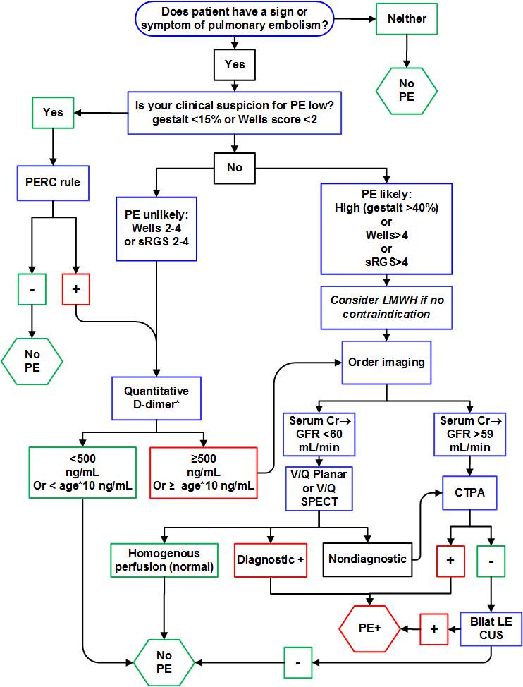
Start work up for PE if patient has any sign (Unexplained tachycardia/tachypnea, low SpO2) or symptom (Chest Pain, SOB) of PE. It is reasonable to consider PE as a differential when ECG shows evidence of right Heart Strain (S1Q3T3, Simultaneous TWI in inferior an pre-cordial leads). 

Caveat: All patients coming with Chest pain do not need a work up for PE. Risk Stratify them and score them on Well's criteria and PERC rule before starting the work up. Random ordering of d-dimer leads to false positive and unnecessary imaging/anticoagulation. 


Random d-dimer sent on a patient with Non-Specific Chest Pain. Can't we just ignore that?
D-Dimer is a great test if used sensibly. It is reassuring if negative (in low risk patients) to exclude PE. Always utilise d-dimer with clinical probability. 


If you are thinking high risk for PE, do not even bother sending a d-dimer. You will need imaging to rule out PE in high-risk regardless of the d-dimer results. As with every test in medicine, there are false positives and false negatives with d-dimer as well. We often talk about the non-specificity of d-dimer and false positives. Here is a list of things that cause false positive and false negative d-dimer.

Of course, you can disregard an elevated d-dimer but you should have a reasonable explanation for that. It should be documented clearly why imaging (CTPA or VQ) was not pursued if d-dimer was found elevated. 


When do we consider thrombolysis for PE?
PEs can be classified as:
  1. Massive PE is defined as acute PE with obstructive shock or SBP <90 mmHg
  2. Submassive PE is acute PE without systemic hypotension (SBP ≥90 mm Hg) but with either RV dysfunction or myocardial necrosis (positive troponin or ECHO evidence of RV dysfunction)
  3. Low risk PEs
Massive PE are definite ones for thrombolysis whereas thrombolysing Submissive PEs is a bit more controversial. Low risk PEs get anti coagulated with heparin. Other indications for using thrombolytics can be Cardiac Arrest with known PE patient or in someone with Presumed PE. 

Click here to read more about thrombolysis in PE. 


Troponin is not helpful in the diagnosis of PE, but it is helpful in the assessment for severity of disease.  


Do we need to treat small distal clots but without DVT (single clot <3mm)?
Isolated distal clots can represent a possible artefact rather than true disease. Many Sub segmental PEs are not even seen by another radiologist when blinded. But guidelines still recommend to treat if risk factors present or if symptomatic and has low risk of bleeding. 


What if CTPA turns negative but you have extremely high suspicion for PE?
Ask radiologist about the quality of scanner. Clinical context holds above everything else. Remember, Gold Standard for diagnosis is Pulmonary Angiography. 


When to consider IVC filter, cather directed lysis?
The indications for IVC filter include: 
(1) Patients with contraindications to anticoagulation
(2) Those who have complications from the use of anti-coagulation
(3) Those who fail to attain adequate anticoagulation while undergoing treatment. 


Cather directed lysis should be first line if available for massive PE. 


Working up PE in pregnancy? 
Start with lower extremity doppler (if DVT+ and hemodynamically stable, start anti-coagulation). Some authorities do not even recommend sending a d-dimer in pregnancy while others recommend using trimester adjusted d-dimer. When used in pregnancy, d-dimer can be extremely helpful when negative. Here is an algorithm based on using trimester adjusted d-dimer.


If you do not want to use d-dimer at all then you have these options:
  • Treat with Heparin Doppler if positive for DVT
  • ECHO/Troponin to further risk stratify
  • CTPA (Radiation is 4 times more than that of a V/Q scan)
  • VQ Scan (done only if CXR is normal, if CXR abnormal then go for CTPA)
             
                                         CTPA - Good for baby, Bad for Mom
                                         V/Q - Bad for baby, Good for Mom


Consider V/Q in cases of Allergy to iodine contrast, Impaired Renal Function and Pregnancy. Issues with V/Q scan can be availability, expertise to interpret the scan. V/Q often gets interpreted as low probability or intermediate probability which leaves you nowhere! 
Too many CTPAs can give you false positives and lead to over-diagnosis and over treatment. Ask yourself, if that tiny clot is really responsible for your patient's symptoms or was it just an incidetaloma! 

The least desirable scenario is one in which both a V/Q scan and a CTPA are needed to complete the diagnostic evaluation. 


Working up PE in a patient already on oral anticoagulants?
Anticoagulants can make d-dimer unreliable and there is no clear consensus on this. 
CTPA them if you have any concerns for a PE and plan for an IVC filter if they are CTPA positive. 


Consider targeting higher INR as they are throwing clots even on oral anti-coagulants. 


Summary:
The testing to catch Pulmonary Embolisms has skyrocketed over the last decade. The increased use of CT in patients with suspected PE has resulted in an increase in the diagnosis of PE but without an associated mortality benefit. Start a work up for PE based on your gestalt or Well's Criteria followed by PERC rule (Low risk on Well's Criteria and PERC negative does not even need a d-dimer). Every test comes with caveats (d-dimer, CTPA, VQ scan) and we must act in a prudent manner while ordering imaging to avoid unnecessary anti-coagulation. 


Further Reading
  • Age-Adjusted d-dimer
  • Tichauer M. The Emergency Medicine Approach To The Evaluation And Treatment Of Pulmonary Embolism. Emergency Medicine Practice. 2012:2.
  • Kline JA, Kabrhel C. Emergency evaluation for pulmonary embolism, Part 2: diagnostic approach. The Journal of emergency medicine. 2015 Jul 31;49(1):104-17.


Posted by:

              
     Lakshay Chanana
     
     Speciality Doctor
     Northwick Park Hospital
     Department of Emergency Medicine
     England
     @EMDidactic


Monday, May 15, 2017

Interpreting Elevated Troponins and Types of Myocardial Infarction

Elevations of cardiac biomarkers in the blood reflect injury leading to necrosis of myocardial cells but biomarkers do not indicate the underlying mechanism of this injury. Myocardial injury can be due to ischemic and non-ischemic causes. Therefore, small amounts of myocardial injury with necrosis may be detected, which are associated with CCF, renal failure, myocarditis, arrhythmias, pulmonary embolism and percutaneous or surgical coronary procedures. Troponin elevation in these contexts should not be called as Myocardial Infarction but Myocardial Injury.


Onset of myocardial ischaemia is the initial step resulting from an imbalance between oxygen supply and demand. This can be identified from history and ECG. Possible ischaemic symptoms include various combinations of chest, upper extremity, mandibular or epigastric discomfort or an ischaemic equivalent such as dyspnoea or fatigue (in women). Pain associated with vomiting, worse on exertion, sweating is definitely worrisome. The discomfort with acute MI usually lasts 20 min. In an ideal world, the sequence should be history, physical exam, ECG and troponins. Whenever symptoms recur, it is important to repeat the ECG. If you are ordering trop troponins, you must order serial ECGs as well. remember, ECG is more of a rule in test not a rule out test for ACS.

MI is classified into various types, based on pathological, clinical and prognostic differences, along with different treatment strategies 


References:

  1. Roe MT, Harrington RA, Prosper DM, Pieper KS, Bhatt DL, Lincoff AM, Simoons ML, Akkerhuis M, Ohman EM, Kitt MM, Vahanian A, Ruzyllo W, Karsch K, Califf RM, Topol EJ. Clinical and therapeutic profile of patients pre- senting with acute coronary syndromes who do not have significant coronary artery disease. The Platelet glycoprotein IIb/IIIa in Unstable angina: Receptor Suppression Using Integrilin Therapy (PURSUIT) trial Investigators. Circulation. 2000;102:1101 – 1106.
  2. Bugiardini R, Manfrini O, De Ferrari GM. Unanswered questions for manage- ment of acute coronary syndrome: risk stratification of patients with minimal disease or normal findings on coronary angiography. Arch Intern Med 2006; 166:1391 – 1395.
  3. Reynolds HR, Srichai MB, Iqbal SN, Slater JN, Mancini GB, Feit F, Pena-Sing I, Axel L, Attubato MJ, Yatskar L, Kalhorn RT, Wood DA, Lobach IV, Hochman JS. Mechanisms of myocardial infarction in women without angiogra- phically obstructive coronary artery disease. Circulation 2011;124:1414 – 1425. 
  4. Bertrand ME, LaBlanche JM, Tilmant PY, Thieuleux FA, Delforge MR, Carre AG, Asseman P, Berzin B, Libersa C, Laurent JM. Frequency of provoked coronary arterial spasm in 1089 consecutive patients undergoing coronary arteriography. Circulation 1982;65:1299 – 1306.
  5. Suwaidi JA, Hamasaki S, Higano ST, Nishimura RA, Holmes DR Jr, Lerman A. Long-term follow-up of patients with mild coronary artery disease and endothe- lial dysfunction. Circulation 2000;101:948 – 954.
  6. Bugiardini R, Manfrini O, Pizzi C, Fontana F, Morgagni G. Endothelial function predicts future development of coronary artery disease: a study on women with chest pain and normal angiograms. Circulation 2004;109:2518 – 2523. 
  7. Kristian Thygesen, Joseph S. Alpert, Allan S. Jaffe, Maarten L. Simoons, Bernard R. Chaitman and Harvey D. White: the Writing Group on behalf of the Joint ESC/ACCF/AHA/WHF Task Force for the Universal Definition of Myocardial Infarction 
Posted by:


     Lakshay Chanana
     
     Speciality Doctor
     Northwick Park Hospital
     Department of Emergency Medicine
     England

     @EMDidactic