Showing posts with label Cardiac Arrest. Show all posts

Wednesday, December 5, 2018

Surviving OHCA by Prof. Richard Lyon

This is the talk by Prof. Richard Lyon at The Bick Sick in Zermatt, 2018


Dr. Lyon is a consultant in Emergency Medicine and clinical lead for Medic1 at the Royal Infirmary of Edinburgh. He is the associate medical director of Kent, Surrey & Sussex Air Ambulance and chair or Pre-hospital Emergency Care at the University of Surrey. He completed a unique doctorate thesis on out-of-hospital cardiac arrest (OHCA) – the TOPCAT study, which has formed the basis of a successful programme of work to improve outcome from OHCA across Scotland. He has won numerous national and international awards for his work. 


Posted by:


     Lakshay Chanana
     
     ST4 Trainee
     Royal Infirmary of Edinburgh
     Department of Emergency Medicine
     Edinburgh
     Scotland

     @EMDidactic

Monday, November 19, 2018

Hypothermic Arrest

Management of accidental hypothermia has seen a revolution with the use of ECLS. ECLS has substantially improved survival and is the treatment of choice in the patient with unstable circulation or CA.The crucial factor in all hypothermia cases is whether critical brain hypoxia occurs before protective brain-cooling takes place


Brain oxygen-consumption decreases by ~6 % per 1 °C fall in core temperature and reaches 16 % at 15 °C compared with normothermia. This improves the brain’s tolerance for low- or no blood-flow states. 


At 18 °C the brain tolerates CA for up to 10 times longer than at 37 °C 


Staging of Hypothermia


Factors affecting the outcome of a hypothermic arrest
(1) hypoxia (the most important single factor)
(2) patient considerations (e.g. age, co-morbidities, trauma)
(3) speed of cooling
(4) environment (air, water, snow)
(5) CA features (body temperature; whether hypoxia preceded arrest; delay before instituting CPR, and CPR quality)
(6) rescue considerations (e.g. adequate training to manage a low flow or no blood-flow state; speed of hospital transfer)
(7) proximity of appropriate hospital facilities
(8) whether hospital staff appreciate the special requirements of these cases


Effects of Hypothermia


Warming Methods


CPR and Drugs
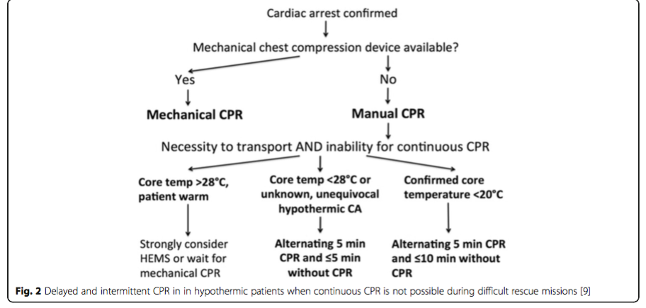
Mechanical chest compression devices (deliver >50 % of baseline cerebral blood flow in normothermia) are likely to provide sufficient oxygen delivery to vital organs in deeply hypothermic patients. When mechanical CPR is not available and manual CPR is not feasible, intermittent >CPR has been suggested. 


International guidance differ on drug administration. ERC 2015 guidelines recommend withholding adrenaline administration in hypothermic CA (HT IV) and limiting defibrillation to three attempts until the core temperature is >30 °C. In contrast, American Heart Association guidelines allow further defibrillation attempts concurrent with rewarming strategies and state that it may be reasonable to consider adrenaline administration during CA according to the standard ALS algorithm.


The benefit of antiarrhythmic drugs in hypothermic CA is unclear. Many arrhythmias (e.g. bradycardia, atrioventricular blocks, atrial fibrillation, nodal rhythms and QRS prolongation with or without Osborn J-waves) are considered benign in accidental hypothermia, usually regress with patient rewarming and do not require further treatment provided the perfusion is deemed adequate. Most intravenous anaesthetic induction agents cause cardiovascular depression so doses should be small. Hypothermia reduces the systemic clearance of CYP450- metabolised drugs by an amount proportional to the fall in body temperature, increasing the likelihood of unanticipated toxicity.


Treatment Algorithm based on Staging and cardiopulmonary status 


Further Reading
Paal P, Gordon L, Strapazzon G, Maeder MB, Putzer G, Walpoth B, Wanscher M, Brown D, Holzer M, Broessner G, Brugger H. Accidental hypothermia–an update. Scandinavian journal of trauma, resuscitation and emergency medicine. 2016 Dec;24(1):111.


Posted by:


     Lakshay Chanana
     
     ST4 Trainee
     Royal Infirmary of Edinburgh
     Department of Emergency Medicine
     Edinburgh
     Scotland

     @EMDidactic

Monday, July 3, 2017

When to stay away from Amiodarone

This week, listen to Amal Mattu talking about Amiodarone. 

Does Amiodarone really fix everything?


Monday, October 19, 2015

Capnography beyond ROSC

What is Capnography?
It is a non-invasive technique where CO2 is analysed continuously, in the gases entering and leaving the lungs. Look at it like a non-invasive way of estimating the PaCO2. Normal ETCO2 ranges between 35-45mmHg.

Different ways of estimating ETCO2:
1. Waveform capnography (Quantitative): When ETCO2 is graphically displayed and analysed quantitatively (real time quantitative waveform as depicted below). Capnograph is the device that measures and displays the waveform. It gives us a number i.e very close to the PaCO2 (typically underestimates PaCO2 by 5mmHg in healthy adults). Therefore, a high ETCO2 is almost always associated with hypercapnia. 

Normal Capnograph (Adults)


2. Capnometry (Quantitative): When only the numerical value is displayed without the waveform.


3. Qualitative: Does not tell us one fixed number instead gives a range of ETCO2 using the calorimetric device. Ex. 21-30mmHg. Hence, utility is limited to ET tube placement verification.

Calorimeter device

How is it different from SpO2?
SpO2 measures O2 saturation in the blood whereas ETCO2 measures CO2 in the airway. ETCO2 reflects the elimination of CO2 by the lungs and thus paints an immediate picture of patient's condition on the monitor, in contrast to SpO2 that remains normal for several minutes (even when you hold your breath).

SpO2 and ETCO2                             
When interpreting ETCO2, look at both, the numeric value as well as the waveform. If you look only at the numeric value, this is like only looking at the heart rate without the ECG wave pattern!


Normal ETCO2 waveform:

Phase I aka Dead space ventilation/ Inspiratory baseline: Beginning of exhalation, no gas exchange here. There is almost no CO2 in airway during inspiration, so baseline is normally zero.

Phase II aka Ascending/Expiratory upstroke/ Early exhalation phase: CO2 from alveoli reaches the upper airway and causes a rapid rise in the amount of CO2 that is detected on the monitor.

Phase III aka Expiratory plateau phase: CO2 rich air is exhaled out
ETCO2 i.e. end of exhalation contains the highest concentration of CO2 (normal value is about 35-45 mmHg).

Phase IV aka Descending/ Expiratory downstroke: Inspiration begins and CO2 in the airways drops down to zero.


Normal capnograph wave
Note: The baseline represents the inspiratory phase (not expiratory)

Normal capnograph
Increase in ETCO2 (See the waveform below)
Hypoventilation
Increased muscular activity
Malignant hyperthermia
Increased Cardiac Output or Bicarbonate infusion
Relief from bronchospasm

Increasing ETCO2 (Ex. Hypoventilation during procedural sedation)


Decrease in ETCO2 (See the waveform below)
Hyperventilation
Muscle relaxants and hypothermia
Decrease cardiac output
Pulmonary embolism 
Bronchospasm

Decreasing ETCO2

Clinical Applications:
  1. Airway management: To confirm the ET tube placement, also for continuous confirmation of the airway
  2. Monitoring CPR efficacy: Target ETCO2 >20 during CPR
  3. Estimates prognosis during CPR: An end-tidal CO2 value < 10 mm Hg after 20 min of resuscitation has been shown to be very accurate in predicting death
  4. Confirming ROSC without stopping chest compressions: With ROSC ETCO2 shows as a sudden increase
  5. Monitoring PaCo2 in a Traumatic Brain Injury/ Post Cardiac Arrest care
  6. Guide to ventilation during procedural sedation: Shows hypoventilation i.e increasing ETCO2 at least 60 seconds prior to hypoxia 
  7. Fluid responsiveness (5% or greater increase in ETCO2 following a passive leg raise is a non invasive way of predicting fluid responsiveness)
  8. Diagnosing other pathologies (PE, DKA)

ETCO2 waveform in other pathologies:

Airway obstruction pattern (Bronchospasm)

Rebreathing

Esophageal intubation

On Muscle relaxants

Take Home
  • ETCO2 is a surrogate marker for arterial CO2. If increased, it confirms hypercapnia.
  • ETCO2 changes immediately with changes in the amount of CO2 in the airway (unlike SpO2 that shows a lag)
  • When using waveform capnography, look at the ETCO2 numeric value as well as the waveform.
  • Eliminate pulse checks during CPR, use ETCO2 instead.

References:
  1. Heradstveit BE, Heltne JK. PQRST - A unique aide-memoire for capnography interpretation during cardiac arrest. Resuscitation 2014; 85:1619-20.
  2. Monnet, X. et al. ETCO2 is better than arterial pressure for predicting volume responsiveness by PLRT. Intensive Care Med. 2013 Jan;39 (1): 93-100
  3. http://www.medscape.com/viewarticle/812011
  4. http://edtech2.boisestate.edu/meganjacobson/502/capno.html
  5. http://www.carefusion.com/pdf/Center_for_Safety/Documents/RC1706-L3017-Capnography-Handbook.final.pdf

Monday, October 12, 2015

Running the code

This week, lets go through some key points on cardiac arrest. Some of these form the fundamentals of CPR in addition to other important points which are not talked about when we do the routine life support courses.

1. High quality CPR
Well, we all know this, Right?
  • Rate:100-120/min
  • Depth: 5cm approximately
  • Allowing full chest recoil
  • Not to hyperventilate
  • Minimising interruptions

This is something that is really stressed on during the life support courses. Well, this is because survival is linked to the quality of CPR and there should not be any excuses here. This is very basic simple stuff that can make a difference. So how are doing it so far?

Current literature says we are slow/ shallow with chest compressions and do not allow adequate recoil. So next time when you run a code, make sure that you stick to these points and especially stay away from interruptions.

Reasons for interruptions: 

a) Airway: Securing the airway should be individualised for every patient. There is no magic number here. For a witnessed arrest with presumed cardiac cause, airway can be delayed for 8-10 minutes whereas early airway control should be done for a hypoxic arrest (drowning, pulmonary edema). In India, pre-hospital intubations are rare and by the time patients are brought to a hospital, airway should be secured ASAP on arrival with minimal interruptions to chest compressions. This is not the time for a novice to try intubation, it should be done by the most experienced provider around. 

b) Pulse Checks: Checking pulse during CPR is unreliable and should not be done, instead use ETCO2 to look for the ROSC. DO NOT STOP COMPRESSIONS TO CHECK FOR PULSE. 

c) Peri shock pauses: Shock can be delivered with transcutaneous pads or paddles and in India, the use of paddles is far more common than pads. 
With every 5 seconds of peri shock pause, there is an 18% decrease in survival. It is recommended to continue to deliver compressions while charging the defibrillator and once it is charged, only then hold the chest compressions for probably < 5 seconds, deliver the shock and resume compressions. It is painful to see those providers, who stop compressions as soon as they touch the paddles, then charge the defibrillator and then deliver the shock. This way it roughly takes about 20-30 seconds.

If there is expertise available, use intra arrest ECHO to pick up a tamponade or dilated RV, but again minimise compressions (Transesophageal ECHO is coming to the ED's soon..)


2. Hemodynamics Guided Resuscitation
Choose one of these three to judge your performance and hemodynamic status:

a) Coronary Perfusion Pressure (needs an arterial line and central line to monitor CPP)
  • Our goal is to achieve an adequate Coronary Perfusion Pressure (CPP) i.e. CPP > 20mm Hg
  • CPP = Aortic DBP - Right Atrial Pressure (RAP) 
OR

b) Diastolic Blood Pressure (needs just an arterial line)
  • Insert a intra arrest femoral arterial line and target DBP > 40mm Hg 
  • If < 40, improve CPR or give epinephrine
  • If > 40, no need to give epinephrine, continue high quality CPR
OR

c) ETCO2 
  • When you can't get an arterial line, use waveform capnography
  • It is the continuous non invasive measurement end tidal CO2 using a sensor, the value is displayed on the monitor as a number.
  • ETCO2 acts as a surrogate marker of cardiac output in addition to confirming the ET tube placement and ROSC.
  • Target ETCO2 > 20mmHg 

3. Medications
Theoretical benefit: Improve CPP, CPP
Detrimental effects: Increase myocardial O2 demand, post ROSC myocardial dysfunction

Epinephrine (Adrenaline)
  • No difference in outcomes, but we are finding it hard to stop using it!!
  • Literature says more epi = no change or worse outcomes.
  • With epi you are more likely to achieve ROSC, but worsen the neurological outcomes.
  • Read more on epinephrine here by Dr. Anand Swaminathan (@EMSwami)
Give q5min epi now if you have been doing q3min so far and watch out for the next ACLS update OR use Hemodynamics Guided Resuscitation. 


3. Team Leadership
  • This is by far the most important take home point from this post. Your leadership skills, ability to mobilise resources and getting things done can affect the outcome of a code. With experience we need to learn how to take control of the situation and communicate effectively under stress. Just knowing the algorithms is not enough.
  • Good Leadership: Appropriate role assignment, better communication, reduces errors and establishes ROSC faster
  • Errors are made due to indecisive and weak leadership which can cost a life.
  • If there is time available, brief your team prior to the code and always debrief after you are done with the code regardless of the outcome. This will improve the team dynamics for future resuscitations. 


So take the charge, assign tasks, be decisive, communicate early, clearly and effectively.

    Take Home:
    • Don't forget the basics, focus on high quality CPR
    • Don't flood them with epinephrine instead use Hemodynamics guided resuscitation (CPP, DBP or ETCO2)
    • Communicate early, clearly and effectively. Good leadership saves lives..

    Thank You


    References:
    1. Michael Winters, MD: Running the Perfect Code in 2015 (AAEM Scientific Assembly) 
    2. Castelao, Ezequiel Fernandez, et al. "Effects of team coordination during cardiopulmonary resuscitation: A systematic review of the literature." Journal of critical care 28.4 (2013): 504-521.CPR quality- Consensus 
    3. Panesar, Sukhmeet S., Agnieszka M. Ignatowicz, and Liam J. Donaldson. "Errors in the management of cardiac arrests: An observational study of patient safety incidents in England." Resuscitation 85.12 (2014): 1759-1763.
    4. Cheskes, Sheldon, et al. "Perishock Pause An Independent Predictor of Survival From Out-of-Hospital Shockable Cardiac Arrest." Circulation 124.1 (2011): 58-66.
    5. Sunde, Kjetil, and Theresa M. Olasveengen. "Towards cardiopulmonary resuscitation without vasoactive drugs." Current opinion in critical care 20.3 (2014): 234-241.
    6. Stiell, Ian G., et al. "Advanced cardiac life support in out-of-hospital cardiac arrest." New England Journal of Medicine 351.7 (2004): 647-656.Johnson NJ et al Resuscitation 2014
    7. Sutton, Robert M., et al. "Hemodynamic-directed cardiopulmonary resuscitation during in-hospital cardiac arrest." Resuscitation 85.8 (2014): 983-986.